| While the technique of expansion microscopy (ExM) is commonly used in animal cells and tissues, plant-specific protocols are very few. Agrisera's collaboration partner, Dr. Kirk Czymmek (Danforth Plant Science Center, USA) and his colleagues from several other American institutions, recently published a method that allows nonoscale imaging by physical expansion of fixed plant protoplasts in a swellable hydrogel. This method, called ExPOSE, leads to enhancement of the resolution by several folds, and shows that, combined with other imagining techniques, like immunofluorescence and in situ hybridization chain reaction, it can lead to visualization of proteins and mRNAs with enhanced spatial resolution. ExPOSE opens new avenues for discoveries in studies of plant cells on the molecular level, as it circumvents the resolution limitations of traditional microscopy methods, and allows the revealing of finer details of cellular structures. Read the recently published article "ExPOSE: a comprehensive toolkit to perform expansion microscopy in plant protoplast systems". Cited Agrisera antibodies: • Anti-GDC-H | H protein of glycine decarboxylase complex (GDC) (AS05 074) • Anti-ACT | Actin (polyclonal) (AS13 2640) • Donkey anti-Rabbit IgG (H&L), DyLight® 594 conjugated, pre-adsorbed (AS12 2076) | 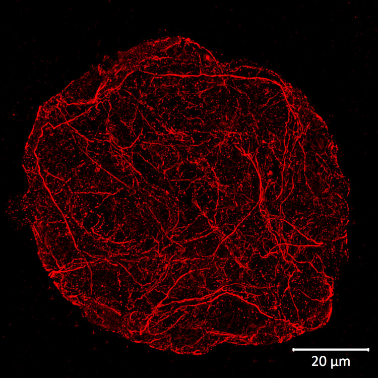 Agrisera polyclonal antibody to plant actin (AS13 2640) was applied to visualize actin filaments using expansion microscopy (ExM) on Arabidopsis thaliana protoplasts. Details of the method are described here. |
Agrisera News
Latest
Interview with Prof. Eiji Nambara2025-04-16 Agrisera anti-Actin antibody applied in a new technique termed Expansion microscopy in plant PrOtoplast SystEms (ExPOSE)
2025-04-11 March and April conferences supported by Agrisera
2025-04-11 Interview with Prof. Rossana Henriques
2025-04-02 New antibody: Anti-FTIP3/FTIP4 | FT Interacting Protein 3/4
2025-04-01 Interview with Prof. Martha Ludwig
2025-03-19 Agrisera supports Annual Congress of Young Researchers (ACYR 2025)
2025-03-14 Interview with Prof. Rebecca Roston
2025-03-05 Agrisera Western blot workshop part I with Yao-Hong Biotechnology for researchers in Taiwan
2025-02-28 Interview with Prof. Junpeng Zhan
2025-02-19
Archive
- April - 2025
- March - 2025
- February - 2025
- January - 2025
- December - 2024
- November - 2024
- October - 2024
- September - 2024
- August - 2024
- July - 2024
- June - 2024
- May - 2024
- April - 2024
- March - 2024
- February - 2024
- January - 2024
- December - 2023
- November - 2023
- October - 2023
- September - 2023
- August - 2023
- July - 2023
- June - 2023
- May - 2023
- April - 2023
- March - 2023
- February - 2023
- January - 2023
- December - 2022
- November - 2022
- October - 2022
- September - 2022
- August - 2022
- July - 2022
- June - 2022
- May - 2022
- April - 2022
- March - 2022
- February - 2022
- January - 2022
- December - 2021
- November - 2021
- October - 2021
- September - 2021
- August - 2021
- July - 2021
- June - 2021
- May - 2021
- April - 2021
- March - 2021
- February - 2021
- January - 2021
- December - 2020
- November - 2020
- October - 2020
- September - 2020
- August - 2020
- July - 2020
- June - 2020
- May - 2020
- April - 2020
- March - 2020
- February - 2020
- January - 2020
- December - 2019
- November - 2019
- October - 2019
- September - 2019
- August - 2019
- July - 2019
- June - 2019
- May - 2019
- April - 2019
- March - 2019
- February - 2019
- January - 2019
- December - 2018
- November - 2018
- October - 2018
- September - 2018
- August - 2018
- July - 2018
- June - 2018
- May - 2018
- April - 2018
- March - 2018
- February - 2018
- January - 2018
- December - 2017
- November - 2017
- October - 2017
- September - 2017
- August - 2017
- July - 2017
- June - 2017
- April - 2017
- March - 2017
- February - 2017
- December - 2016
- November - 2016
- October - 2016
- September - 2016
- August - 2016
- July - 2016
- June - 2016
- May - 2016
- April - 2016
- March - 2016
- February - 2016
- January - 2016
- December - 2015
- November - 2015
- October - 2015
- September - 2015
- August - 2015
- July - 2015
- June - 2015
- May - 2015
- March - 2015
- February - 2015
- January - 2015
- December - 2014
- November - 2014
- October - 2014
- September - 2014
- August - 2014
- July - 2014
- June - 2014
- May - 2014
- April - 2014
- March - 2014
- February - 2014
- January - 2014
- December - 2013
- November - 2013
- September - 2013
- August - 2013
- July - 2013
- June - 2013
- May - 2013
- April - 2013
- February - 2013
- January - 2013
- December - 2012
- October - 2012
- September - 2012
- August - 2012
- July - 2012
- June - 2012
- May - 2012
- April - 2012
- March - 2012
- December - 2011
- November - 2011
- September - 2011
- August - 2011
- July - 2011
- April - 2011
- January - 2011
- December - 2010
- October - 2010
- September - 2010
- August - 2010
- July - 2010
- March - 2010
- January - 2010
- December - 2009
- November - 2009
- September - 2009
- July - 2009
- June - 2009
- May - 2009
- March - 2009
- January - 2009
- December - 2008