1

Anti-PTOX | Plastid terminal oxidase
AS23 4895 | Clonality: Polyclonal| Host: Rabbit | Reactivity: Arabidopsis thaliana, Chlamydomonas reinhardtii
- Product Info
-
Immunogen: Recombinant PTOX of Arabidopsis thaliana, expressed in E.coli, UniProt: Q56X5, TAIR:AT4G22260 Host: Rabbit Clonality: Polyclonal Purity: Antigen affinity purified serum, in PBS pH 7.4 Format: Lyophilized Quantity: 50 µg Reconstitution: For reconstitution add 50 µl, of sterile or deionized water. Storage: Store lyophilized/reconstituted at -20°C; once reconstituted make aliquots to avoid repeated freeze-thaw cycles. Please, remember to spin tubes briefly prior to opening them to avoid any losses that might occur from lyophilized material adhering to the cap or sides of the tubes. Tested applications: Western blot (WB) Recommended dilution: 1 : 2000 - 1: 5000 (WB) Expected | apparent MW: 40.5 | 34.3 kDa (due to N-terminal or C-terminal processing) - Reactivity
-
Confirmed reactivity: Arabidopsis thaliana, Chlamydomonas reinhardtii Predicted reactivity: Camelina sativa, Brassica napus, Capsella rubella, Capsicum annuum, Citrus sinensis, Cucumis sativus, Eutrema salsugineumm, Glycine max, Gossypium hirsutum, Malus domestica, Medicago truncalula, Nicotiana tabacum, Oryza sativa Japonica Group, Physcomitrium patens, Populus alba x Populus x berolinensis, Pisum sativum, Pistacia vera, Ricinus communis, Solanum lycopersicum, Spinacia oleracea, Sorghum bicolor, Vitis vinifera
Species of your interest not listed? Contact us - Application Examples
-

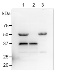
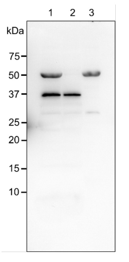
Samples:
1 - Chloroplast samples corresponding to 3.0 µg chlorophyll of Arabidopsis thaliana
2 – Thylakoid membrane samples corresponding to 3.0 µg chlorophyll of Arabidopsis thaliana
3 – Stromal fraction samples corresponding to 3.0 µg chlorophyll of Arabidopsis thaliana
Left panel: MW markersProtein samples corresponding to 1.0 µg chlorophyll of chloroplasts extracted from Arabidopsis thaliana. Chloroplast samples were isolated with 20 mM Tricine–NaOH, pH 8.4, containing 400 mM sorbitol, 5 mM MgCl2, 5 mM MnCl2, 2 mM EDTA, 10 mM NaHCO3, 0.5% (w/v) bovine serum albumin (BSA) and 5 mM ascorbate: and denatured with exact buffer (62.5 mM Tris-HCl, pH 6.8, containing 2% (w/v) SDS, 10% (w/v) glycerol, 0.005% (w/v) bromophenol blue, and 50 mM dithiothreitol) at 95 °C/5 min. Samples were separated at RT on 15% acrylamide gel and blotted for 0.5 h to PVDF (pore size of 0.2 µm), using: semi-dry transfer at RT. Blot was blocked with 5 % milk for: 1h/RT with agitation. The blot was incubated in the primary antibody at a dilution of 1: 2,000 for 1h/RT with agitation in TBS-T. The antibody solution was decanted, and the blot was rinsed briefly twice, then washed 3 times for 5 min in TBS-T at RT with agitation. Blot was incubated in matching secondary antibody (AS09 602 anti-rabbit IgG horse radish peroxidase conjugated) diluted to 1: 25,000 in for 1h/RT with agitation. The blot was washed as above and developed with a following chemiluminescent detection reagent: AS16 ECL-N-10 AgriseraBright (mid picogram). Exposure time was 3 minutes.
Note: The band at 50 kDa is a cross reactivity with Rubisco.Courtesy of Prof.Yuki Okegawa, Okayama University, Japan

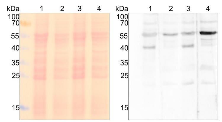
Samples:
l1 - Chlamydomonas reinhardtii, wild type cc124 (mt -)
2 - Chlamydomonas reinhardtii ptox2.34 (mt -)
3 - Chlamydomonas reinhardtii wild type cc125 (mt +)
4 - Chlamydomonas reinhardtii ptox2.24 (mt +)
1x10e6 cells per lane of total protein extracted with Chloroform/Methanol protein precipitation protocol (dx.doi.org/10.17504/protocols.io.icecate) from Chlamydomonas reinhardtii. Exact buffer components were: Count Chlamydomonas reinhardtii cell density in TAP cultures (early log phase), spin down 15 ml culture (4000 g, 5 min, 22°C) and resuspend pellet in 1 ml supernatant. Use 200 µl resuspended cells for protein precipitation in 2 ml Eppendorf tubes at room temperature (dx.doi.org/10.17504/protocols.io.icecate). Pellets were dried from MeOH and resuspended in 1x SDS PAGE running buffer (50 mM Tris/HCl pH 6.8, 50 mM DTT, 50 mM Na2CO3, 2% (w/v) SDS, 12% (v/v) Glycerol, 0.01% (w/v) Bromphenolblau, 0.01% (w/v) Servablau G) so that 20 µl corresponds to 1x10e6 cells. Sample was denatured (20 min, 22 °C) and spun down before loading. 13% SDS-PAGE Laemmli 1 h to nitrocellulose (pore size of 0.45 µm) semi-dry at room temperature. Ponceau S staining of total proteins to check loading and transfer efficiency. Follow recommended protocol for the remaining procedure (use agitation throughout; no double rinsing but in between blocking & antibody changes include 3x washing steps with TBS-T, 5 min/RT). Blocking: 5% non-fat milk 1h/RT, primary anti-PTOX antibody (Product No AS23 4895, Lot 2309) 1:1000 in TBS-T ON/4C, secondary antibody (AS09 602-trial) 1:25000 1h/RT, detection first with Agrisera Bright AS16 ECL-N. Exposure time was 3 s.
Courtesy of Dr. Felix Buchert, University of Munster, Germany - Background
-
Background: PTOX (plastid terminal oxidase) is a component of electron transfer chain responsible for desaturation of phytoene, which prevents the generation of reactive oxygen species. It is involved in the differentiation of plastids: chloroplasts, amyloplasts, and etioplasts. PTOX is expressed ubiquitously in plant tissues and is located in the lumen.Synonymes: IM, IM1, immutants, AOX4, alternative oxidase 4, ubiquinol oxidase 4, chloroplastic/chromoplastic. - Protocols
-
Agrisera Western Blot protocol and video tutorials
Protocols to work with plant and algal protein extracts
Agrisera Educational Poster Collection - Reviews:
-
This product doesn't have any reviews.

