1

Anti-NAI2 | TSA1-like protein (ER lumen marker)
- Product Info
-
Immunogen: Purified recombinant NAI2 of Arabidopsis thaliana, residues 272-502 with a His tag, UniProt: Q9LSB4, TAIR: At3g15950 Host: Rabbit Clonality: Polyclonal Purity: Total IgG. Protein A purified in PBS, 50% glycerol. Filter sterilized. Format: Liquid at 2 mg/ml. Quantity: 200 µg Storage: Store at -20°C; once reconstituted make aliquots to avoid repeated freeze-thaw cycles. Please remember to spin the tubes briefly prior to opening them to avoid any losses that might occur from material adhering to the cap or sides of the tube. Tested applications: Immunofluorescence (IF), Western blot (WB) Recommended dilution: 1: 1000 - 1: 3000 (IF), 1: 2000 - 1: 4000 (WB) Expected | apparent MW: 85 | 120 kDa - Reactivity
-
Confirmed reactivity: Arabidopsis thaliana Predicted reactivity: Species of your interest not listed? Contact us Not reactive in: No confirmed exceptions from predicted reactivity are currently known - Application Examples
-

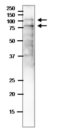
Arabidopsis thaliana 7 day-old seedlings were freshly extracted with 2x SDS-sample buffer (+ 2ME) for SDS-PAGE and denatured with 4X SDS buffer at 95°C for 5 min. Protein load/well is 10 µg. Sample was separated on 12.5 % SDS-PAGE and blotted to PVDF membrane using wet tank system. Blot was blocked with 3 % skim milk/TBS-T, 1h/RT with agitation. Blot was incubated in the primary antibody at a dilution of 1: 4000 in TBS-T for 1h/RT. The antibody solution was decanted and the blot was washed 4 times for 10 min in TBS-T at RT with agitation. Blot was incubated in matching secondary antibody (anti-rabbit IgG horse radish peroxidase conjugated) diluted to 1:10 000 in for 1h/RT with agitation. The blot was washed as above and developed with a chemiluminescent detection reagent, following manufacture's recommendation.
Upper band is NAI2, while the lower band is a degradation product of NAI2 (Yamada et al. 2008).
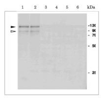
Total protein was extracted from 20 Arabidopsis thaliana 7 day-old seedlings using 200 ml of 20 mM Tris-HCl pH 6.8, 40 % glycerol, 2 % SDS and 2 % ME and denatured with 4X SDS buffer at 95°C for 5 min. Samples was separated on 12.5 % SDS-PAGE and blotted using wet transfer to nitrocellulose membrane. Blot was blocked with 3 % skim milk/TBS-T, 1h/RT with agitation. Blot was incubated in the primary antibody at a dilution of 1: 2000 in TBS-T for 1h/RT. The antibody solution was decanted and the blot was washed 4 times for 10 min in TBS-T at RT with agitation. Blot was incubated in matching secondary antibody (anti-rabbit IgG horse radish peroxidase conjugated) diluted to 1:10 000 in for 1h/RT with agitation. The blot was washed as above and developed with a chemiluminescent detection reagent, following manufacture's recommendation. Samples: Arabidopsis thaliana wild-type (1), Arabidopsis thaliana wild-type with GFP-h (2), mutant nai2-1 (3), mutant nai2-5 (4), mutant nai2-3 (5), mutant nai1-1 (6)
Immunofluorescence staining of NAI peotein in ER bodies.
Immunofluorescence analysis of NAI2 in 7-d-old GFP-h seedlings. Left panels: the ER-targeted GFP signals; middle panel: the NAI2 signals, which were detected by antibodies against NAI2/ΔSP, and Cy3-labeled second antibodies; right panel, the merged images. Bars = 10 mm. - Additional Information
-
Additional information (application): Signal peptide of 24 amino acids is removed from the mature protein - Background
-
Background: NAI2 TSA1-like protein is responsible for the ER body formation. Regulates the number and shape of the ER bodies and the accumulation of PYK10 in ER bodies, but is not involved in the expression of PYK10. Interacts directly or indirectly with MEB1 and MEB2. Expressed in roots. Detected in shoot apex. Induced by NAI1. The protein is localising to ER lumen. - Product Citations
-
Selected references: Ueda et al. (2018).Endoplasmic Reticulum (ER) Membrane Proteins (LUNAPARKs) are Required for Proper Configuration of the Cortical ER Network in Plant Cells. Plant Cell Physiol. 2018 Oct 1;59(10):1931-1941. doi: 10.1093/pcp/pcy137.
Yamada et al. (2013). Identification of two novel endoplasmic reticulum body-specific integral membrane proteins. Plant Physiol. 2013 Jan;161(1):108-20. doi: 10.1104/pp.112.207654.
Yamada et al. (2008). NAI2 is an endoplasmic reticulum body component that enables ER body formation in Arabidopsis thaliana. Plant Cell . 2008 Sep;20(9):2529-40. doi: 10.1105/tpc.108.059345.
- Protocols
-
Agrisera Western Blot protocol and video tutorials
Protocols to work with plant and algal protein extracts
Agrisera Educational Posters Collection - Reviews:
-
This product doesn't have any reviews.
Accessories

AS09 481 | Clonality: Polyclonal | Host: Rabbit | Reactivity: A. thaliana, B. napus, Chara australis, C. reinhardtii, C. sativus, M. perniciosa, N. benthamiana, N. tabacum, R. sativa, L.Tokinashi-daikon, O. europaea, O. sativa, P. abies, P. patens, S. oleracea, S. lycopersicum, S. tuberosum, T. aestivum, Z. mays


