1

Anti-AGO9 | Argonaute 9
AS10 673 | Clonality: Polyclonal | Host: Rabbit | Reactivity: Arabidopsis thaliana
- Product Info
-
Immunogen: KLH-conjugated synthetic peptide derived from Arabidopsis thaliana AGO9 protein sequence UniProt: Q84YI4, TAIR:At5g21150.
Host: Rabbit Clonality: Polyclonal Purity: Immunogen affinity purified serum in PBS pH 7.4. Format: Lyophilized Quantity: 50 µg Reconstitution: For reconstitution add 50 µl of sterile water Storage: Store lyophilized/reconstituted at -20°C; once reconstituted make aliquots to avoid repeated freeze-thaw cycles. Please remember to spin the tubes briefly prior to opening them to avoid any losses that might occur from material adhering to the cap or sides of the tube. Tested applications: Immunolocalization, whole-mount (IL), Immunoprecipitation (IP), Western blot (WB) Recommended dilution: 5 µg of antibody per 1 gram of a fresh tissue (IP), 1 : 10 000 (WB) Expected | apparent MW: 101 kDa
- Reactivity
-
Confirmed reactivity: Arabidopsis thaliana Predicted reactivity: Arabidopsis thaliana
Not reactive in: No confirmed exceptions from predicted reactivity are currently known - Application Examples
-
Application example

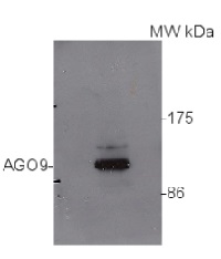
80 µg of Arabidopsis thaliana soluble total cell extract (extracted in 20 mMTris pH7.5, 5mM MgCl2, 2.5mM DTT, 300mM NaCl, 0.1% NP-40, 1% proteaseinhibitor) was separated on 6% SDS-PAGE and blotted 1h to nitrocellulose. Filters were blocked 1h with 5% low-fat milk powder in TBS-TT (0.25% TWEEN20; 0.1% Triton-X) and probed with anti-AGO9 antibody (1:10 000, 1h) and secondary anti-rabbit antibody (HRP conjugated, Agrisera AS09 602) (1:15 000, 1 h) in TBS-TT containing 5% low fat milk powder. Antibody incubations were followed by washings in TBS-TT. All steps were performed at RT with agitation. Blots were developed for 5 min with chemiluminescent detection reagent according the manufacturer's instructions. Exposure time was 30 seconds.
Courtesy Dr. Ericka Havecker, University of Cambridge, UK
- Additional Information
-
Additional information (application): AGO expression may be cell/tissue specific and using floral tissue is recommended where most of the AGOs are expressed the highest. Seedlings can be used as a negative control.
Use of proteasome inhibitors as MG132 can help to stabilize AGO proteins during extraction procedure.
A recommended whole-mount immunolocalization protocol can be found here. - Background
-
Background: AGO9 belongs to a group of argonaute proteins which are catalytic component of the RNA-incudes silencing complex (RISC).
- Product Citations
-
Selected references: Hou et al. (2021) High-throughput single-cell transcriptomics reveals the female germline differentiation trajectory in Arabidopsis thaliana. Commun Biol. 2021 Oct 1;4(1):1149. doi: 10.1038/s42003-021-02676-z. PMID: 34599277; PMCID: PMC8486858. (immunolocalization)
Oliver & Martinez. (2021) Accumulation dynamics of ARGONAUTE proteins during meiosis in Arabidopsis. Plant Reprod. 2021 Nov 23. doi: 10.1007/s00497-021-00434-z. Epub ahead of print. PMID: 34812935.
Sprunck et al. (2019). Elucidating small RNA pathways in Arabidopsis thaliana egg cells. http://dx.doi.org/10.1101/525956
Su et al. (2017). The THO Complex Non-Cell-Autonomously Represses Female Germline Specification through the TAS3-ARF3 Module. Curr Biol. 2017 Jun 5;27(11):1597-1609.e2. doi: 10.1016/j.cub.2017.05.021.
Havecker et al. (2010) The RNA-directed DNA methylation Arabidopsis Argonautes functionally diverge based on expression and interaction with target loci. Plant Cell 22(2): 321-34. - Protocols
-
TCA/Acetone protein precipitation method for plant proteins
- Grind plant tissue in a liquid nitrogen.
- Continue grinding with 10% TCA solution in acetone (ice cold).
- Precipitate overnight in -20C.
- Spin at 4°C for 1min, 17k rpm > wash with ice cold acetone until you obtain a white pellet.
- Dissolve the pellet in buffer of choice (for example 8M urea containing 5mM DTT, or denaturate in SDS protein loading buffer for 10 min. at 70°C)
- Clarify supernatant
- Measure protein concentration.
- Proceed with a western blot.

360 µg/well of Arabidopsis thaliana protein extracted by TCA-acetone precipitation from floral tissue and saturated in 8M urea were separated on 15% SDS-PAGE and blotted for 1hour to 0.2 µm nitrocellulose at 100V using wet transfer system. Blots were blocked with 0.5% cold fish gelatin for 1hr at room temp with agitation. Blot was incubated in the primary antibody at a dilution of 1:2500 for an hour at RT with agitation. The blots were washed with 3X 15min TBS-TT at RT with agitation. Blots as incubated in the secondary antibody (DayLight 800) 1:5000 dilution for 30 min. at RT with agitation and washed 1X with TBSTT for 15 min, 1X with TBST for 15min before scanning with the ODyssey IRD scanner.
Courtesy of Dr. Betty Chung and Pawel Baster, University of Cambridge, United Kingdom
Recommended protocol for whole-mount immunolocalization using anti-AGO9 antibodies can be found here.
BACK to Plant Protocols page
Agrisera Western Blot protocol and video tutorials
Protocols to work with plant and algal protein extracts
Agrisera Educational Posters Collection
- Reviews:
-
This product doesn't have any reviews.
Accessories

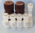
This set contains:
- 4 Anti-AGO antibodies of your choice, chosen from a drop down menu below
- Matching secondary antibody, HRP conjugated, min. 1: 25 000, 1h/RT (AS09 602 - trial)


