1

Anti-TOP2 | DNA topoisomerase II
AS05 096 | Clonality: Polyclonal | Host: Rabbit | Reactivity: Arabidopsis thaliana, Vicia faba
- Product Info
-
Immunogen: The C-terminal 153 amino acids of the Arabidopsis thaliana Topoisomerase II (At3g23890, protein accesion number P30182) with an N-terminal hexahistidine tag was expressed in E.coli and purified by Ni2+ affinity chromatography.
Host: Rabbit Clonality: Polyclonal Purity: Serum Format: Lyophilized Quantity: 200 µl Reconstitution: For reconstitution add 200 µl of sterile water Storage: Store lyophilized/reconstituted at -20°C; once reconstituted make aliquots to avoid repeated freeze-thaw cycles. Please remember to spin the tubes briefly prior to opening them to avoid any losses that might occur from material adhering to the cap or sides of the tube. Tested applications: Immunolocalization (IL), Western blot (WB) Recommended dilution: 1 : 500 (IL), 1 : 2000 (WB) Expected | apparent MW: 164 | 170 kDa (Arabidopsis thaliana)
- Reactivity
-
Confirmed reactivity: Arabidopsis thaliana, Vicia faba Predicted reactivity: Brassica rapa, Chlamydomonas reinhardtii, Chlorella vulgaris, Citrus clementina, Glycine max, Hordeum vulgare, Medicago truncatula, Oryza sativa, Ostreococcus tauri, Panicum italicum, Phaseolus vulgaris, Physcomitrium patens, Pinus sitcHensis, Populus trichocarpa, Solanum tuberosum, Sorghum bicolor, Triticum aestivum, Vitis vinifera, Volvox Caterii
Species of your interest not listed? Contact usNot reactive in: Nicotiana tabacum
- Application Examples
-
Application example
.jpg)
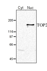
10 µg of total protein from (Cyt) Arabidopsis thaliana cytoplasmic fraction, (Nuc) Arabidopsis thaliana nuclear fraction were separated on SDS-PAGE and blotted 1h to nitrocellulose. Filters were probed with anti-TOP2 antibodies (AS05 096, 1:2000). Signal was detected with chemiluminescent detection reagent.

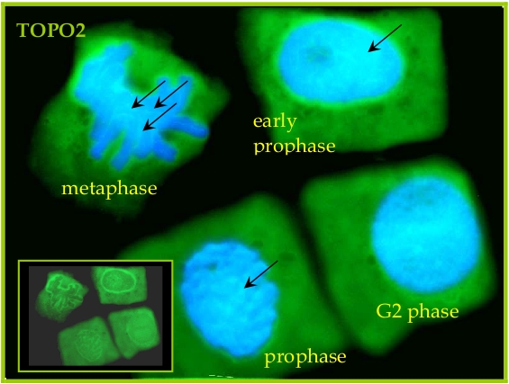
Seeds of field bean (Vicia faba L. subsp. minor var. Nadwiślański; DANKO Group; Sobiejuchy) were sterilized using sodium hypochlorite (0.3% v/v) and germinated in Petri dishes on wetted filter paper at room temperature. At 4 d after imbibition, dark-grown seedlings with primary roots 25±5 mm long were selected for experiments. During incubations roots were oriented horizontally in a humid chamber and aerated continuously on a rotary water-bath shaker (30 rpm) at 23°C. Immunocytochemical assays were performed according to the method prescribed earlier (Rybaczek and Maszewski 2006). Excised apical parts of roots (1.5 mm long) were fixed for 45 min (18°C) in PBS-buffered 3.7% paraformaldehyde, washed several times with PBS and placed in a citric acid-buffered digestion solution (pH 5.0; 37°C for 45 min) containing 2.5% pectinase (Fluka), 2.5% cellulase (Onozuka R-10; Serva) and 2.5% pectoliase (ICN). After removing the digestion solution, root tips were washed 3 times in PBS, rinsed with distilled water and squashed onto Super Frost Plus glass slides (Menzel-Gläser). Air-dried slides were pretreated with PBS-buffered 5% BSA at 20°C for 50 min and incubated overnight in a humidified atmosphere (4°C) with rabbit antibody raised against TOPO2 (Agrisera), dissolved in PBS containing 1% BSA (at a dilution of 1:500). Following incubation, slides were washed 3 times with PBS and incubated for 1 h (18°C) with secondary goat anti-rabbit IgG DyLight®488 antibody (Agrisera, AS09 633, 1:3000). Nuclear DNA was stained with 4’,6-diamidino-2-phenyl-indole (DAPI, 0.4 μg/ml; Sigma-Aldrich). Following washing with PBS, slides were air dried and embedded in Vectashield Mounting Media for Fluorescence (Vector Laboratories). Observations were made using Optiphot-2 fluorescence microscope (Nikon) equipped with B-2A filter (blue light; λ ≈ 495 nm) for DyLight-conjugated antibodies and UV-2A filter (UV light; λ ≈ 365 nm) for DAPI. All images were recorded at exactly the same time of integration using DXM 1200 CCD camera.
Courtesy Dr. Dorota Rybaczek, Lodz University, Poland
- Additional Information
-
Additional information: Antibody detects a protein of ca. 170 kDa on western blots of Arabidopsis thaliana protein extracts. In subcellular fractions of cultured Arabidopsis thaliana cells the antibody detects a 170 kDa protein exclusively in the nulear fraction (see picture).
Additional information (application): Topoisomerase II is highly expressed in young seedlings. The protein is localized in the nucleus and gene expression levels are increased in proliferative tissues like shoot apex or root tip.
This product can be sold containing ProClin if requested. - Background
-
Background: Topoisomerase type II (EC5.99.1.3) is one of the enzymes which is catalyzing unknotting of DNA by creating transient breaks in the DNA using a conserved tyrosine as the catalytic residue.Synonyme names of this protein: At3g23890, ATTOPII, DNA topoisomerase 2, DNA topoisomerase II, F14O13.7, TOP2, TOPOISOMERASE II
- Product Citations
-
Selected references: Martinez-Garcia M et al. (2018). TOPII and chromosome movement help remove interlocks between entangled chromosomes during meiosis. J Cell Biol. 2018 Dec 3;217(12):4070-4079. doi: 10.1083/jcb.201803019.
Xie S & Lam E (1994) Abundance of nuclear DNA topoisomerase II is correlated with proliferation in Arabidopsis thaliana. NAR 25:5729.Klaus Feldmann (1997) Regulation der Topoisomerase II von Arabidopsis thaliana im Zellzyklus. PhD thesis, University of Cologne.
- Protocols
-
Agrisera Western Blot protocol and video tutorials
Protocols to work with plant and algal protein extracts - Reviews:
-
This product doesn't have any reviews.


