1

Anti-RPL1 | 50S ribosomal protein L1
AS11 1738 | Clonality: Polyclonal | Host: Rabbit | Reactivity: cyanobacteria
- Product Info
-
Immunogen: KLH-conjugated synthetic peptide derived from all known cyanobacterial Rpl1 sequences including Synechocystis sp. 6803, P36236
Host: Rabbit Clonality: Polyclonal Purity: Serum Format: Lyophilized Quantity: 50 µl Reconstitution: For reconstitution add 50 µl of sterile water Storage: Store lyophilized/reconstituted at -20°C; once reconstituted make aliquots to avoid repeated freeze-thaw cycles. Please remember to spin the tubes briefly prior to opening them to avoid any losses that might occur from material adhering to the cap or sides of the tube. Tested applications: Western blot (WB) Recommended dilution: 1 : 2000 (WB) Expected | apparent MW: 25 kDa
- Reactivity
-
Confirmed reactivity: Anabaena sp., Nostoc sp., Synechococcus sp. 7942, Synechocystis sp. 6803 Predicted reactivity: Cyanobacteria Not reactive in: E.coli
- Application Examples
-
application example

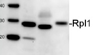
5 µg of total protein from Anabaena sp. (1), Synechococcus sp. 7942 (2), Synechocystis sp. 6803 (3) extracted with Agrisera protein extraction buffer PEB were separated on 4-12% NuPAGE and blotted 1h to PVDF. Blots were blocked with ECL Advance Blocking Reagent for 1h at room temperature (RT) with agitation. Blot was incubated in the primary antibody at a dilution of 1: 10 000 for 1h at RT with agitation. The antibody solution was decanted and the blot was rinsed briefly twice, then washed once for 15 min and 3 times for 5 min in TBS-T at RT with agitation. Blot was incubated in secondary antibody (anti-rabbit IgG horse radish peroxidase conjugated, from Agrisera, AS09 602) diluted to 1:50 000 in for 1h at RT with agitation. The blot was washed as above and developed for 5 min with ECL Advance according to the manufacturers instructions (GE Healthcare). Exposure time was 180 seconds.
Application examples: 
Reactant: Synechocystis
Application: Western Blotting
Pudmed ID: 29985458
Journal: Sci Rep
Figure Number: 2A
Published Date: 2018-07-09
First Author: Koskinen, S., Hakkila, K., et al.
Impact Factor: 4.13
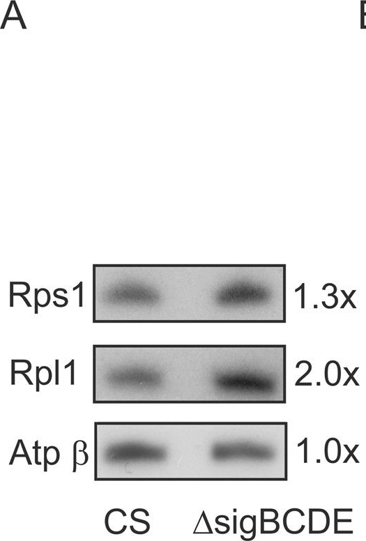
Open PublicationRibosomal proteins, translation activity and total protein contents in the ?sigBCDE and control (CS) strains in standard growth conditions. (A) Total proteins were isolated, separated by SDS-PAGE and then the Rps1 protein of the 30S ribosomal subunit and the Rpl1 protein of the 50S ribosomal subunit were detected with western blotting. As a loading control the amount of the ? subunit of ATPase was detected as well. Original Western blots are shown in Supplementary Fig. S1. (B) Cells were labelled with radioactive 35S-methionine for 10 or 30?min, as indicated, in standard growth conditions, and total proteins were isolated after addition of excess of cold methionine. Proteins were separated by SDS-PAGE, transferred to the membrane and visualized by autoradiography. (C) Total proteins were isolated from the same amount of cells and the protein content was detected with a BioRad DC protein assay kit. Three independent biological replicates were measured, Student’s t-test P?=?0.905.

Reactant: Synechocystis
Application: Western Blotting
Pudmed ID: 29985458
Journal: Sci Rep
Figure Number: 3B
Published Date: 2018-07-09
First Author: Koskinen, S., Hakkila, K., et al.
Impact Factor: 4.13
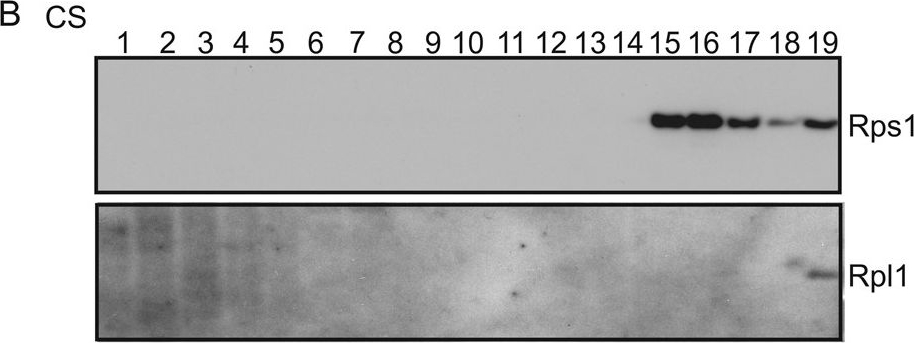
Open PublicationRibosome profiles of the control (CS) and ?sigBCDE strains. The ribosomes of the CS (A,B) and ?sigBCDE (C,D) strains were fractionated with a 5–35% sucrose density gradient centrifugation, nineteen fractions were collected and either RNA or ribosomal proteins Rps1 and Rpl1 were detected from each fraction. RNA was isolated from the 300??L sample of fraction, dissolved in 20??L of water, and 15??L of isolated RNA from each fraction of CS (A) or ?sigBCDE (C) was loaded on a 1.2% agarose gel and stained with ethidium bromide. The DNA molecular marker was included to demonstrate separation of fragments. For western blots, a 24??L sample of a fraction was solubilized and proteins were separated on SDS-PAGE and Rps1 and Rpl1 proteins were detected by specific antibodies by western blotting in the control (B) and ?sigBCDE (D) strains.

Reactant: Synechocystis
Application: Western Blotting
Pudmed ID: 29985458
Journal: Sci Rep
Figure Number: 3D
Published Date: 2018-07-09
First Author: Koskinen, S., Hakkila, K., et al.
Impact Factor: 4.13
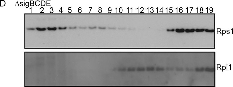
Open PublicationRibosome profiles of the control (CS) and ?sigBCDE strains. The ribosomes of the CS (A,B) and ?sigBCDE (C,D) strains were fractionated with a 5–35% sucrose density gradient centrifugation, nineteen fractions were collected and either RNA or ribosomal proteins Rps1 and Rpl1 were detected from each fraction. RNA was isolated from the 300??L sample of fraction, dissolved in 20??L of water, and 15??L of isolated RNA from each fraction of CS (A) or ?sigBCDE (C) was loaded on a 1.2% agarose gel and stained with ethidium bromide. The DNA molecular marker was included to demonstrate separation of fragments. For western blots, a 24??L sample of a fraction was solubilized and proteins were separated on SDS-PAGE and Rps1 and Rpl1 proteins were detected by specific antibodies by western blotting in the control (B) and ?sigBCDE (D) strains.
- Background
-
Background: Rpl1 (50S ribosomal protein L1) belongs to the ribosomal protein L1P family. This protein binds directly to 23S rRNA, acts also as a translational repressor protein.
- Product Citations
-
Selected references: Brenes-Álvarez et al. (2024). R-DeeP/TripepSVM identifies the RNA-binding OB-fold-like protein PatR as regulator of heterocyst patterning. Nucleic Acids Res. 2024 Dec 19:gkae1247. doi: 10.1093/nar/gkae1247.
Linhartová et al. (2014). Accumulation of the Type IV prepilin triggers degradation of SecY and YidC and inhibits synthesis of Photosystem II proteins in the cyanobacterium Synechocystis PCC 6803. Mol Microbiol. 2014 Jul 24. doi: 10.1111/mmi.12730. - Protocols
-
- Reviews:
-
This product doesn't have any reviews.
Accessories

AS13 2650 | Clonality: Polyclonal | Host: Rabbit | Reactivity: Arabidopsis thaliana, Hordeum vulgare, Nicotiana benthamiana, Zea mays


