1
Anti-RD | N-terminal arginylation
AS15 3050A | Clonality: Polyclonal | Host: Rabbit | Reactivity: RD | N-terminal arginylation
- Product Info
-
Immunogen: KLH-conjugated synthetic peptide: H-RDHKHANQHMSVC-NH2
Host: Rabbit Clonality: Polyclonal Purity: Immunogen affinity purified serum in PBS pH 7.4. Format: Lyophilized Quantity: 2 x 25 µg Reconstitution: For reconstitution add 25 µl of sterile water to each tube Storage: Store lyophilized/reconstituted at -20°C; once reconstituted make aliquots to avoid repeated freeze-thaw cycles. Please remember to spin the tubes briefly prior to opening them to avoid any losses that might occur from material adhering to the cap or sides of the tube. Tested applications: Dot blot (Dot) Recommended dilution: 1 : 1000 (Dot) - Application Examples
-
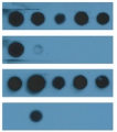
Application information 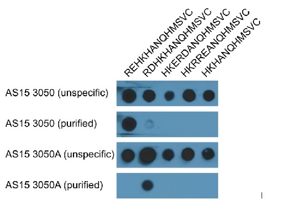
- Membrane: PVDF, Hybond 0.45 μm (GE Healthcare)
- 0.5 µg synthetic peptide per spot
- Membrane was blocked overnight in TBST, 4% Blocking Agent (GE Healthcare)
- Primary antibodies: 1:10 000 in TBST 2% Blocking Agent
- Secondary antibody (AS09 602): 1:25 000 in TBST 2% Blocking Agent
- Detection: ECL
Courtesy of Dr. Sebastian Hoernstein, Faculty of Biology, University of Freiburg; Germany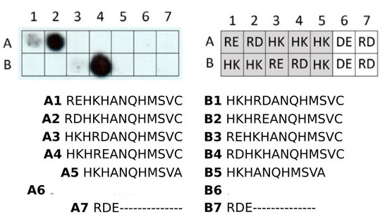
Dotblot of peptide variants probed with anti-RD antibodies1. The respective volumes of a 0.1 mM aqueous peptide solution were mixed with each 1 µl of 0.1% SDS in 0.5 M Na-PO4 (according to Canas et al. 1993, Anal Biochem. 211), dried in a vacuum concentrator and reconstituted in 1 µl of H2O. The peptide solutions were spotted onto a PVDF membrane and air-dried overnight. The dried membrane was subjected to semi-dry western blot electrotransfer (0.85 mA/cm2, 3 min), blocked with 4% skim milk powder in TBST overnight and probed with anti-RD antibody (at 0.4 µg/ml in TBST) and subsequently with HRP coupled anti-sheep antibody. Detection with ECL substrate of femtogram sensitivity, 3 min exposure time.
Courtesy Dr. Nico Dissmeyer, Leibniz Institute of Plant Biochemistry (IPB), Germany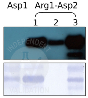
SPOT assay with anti-RD antibody. 11- to 17mer peptides of the indicated sequences were synthesized on a PEG-derivatized cellulose membrane. The SPOT membrane was blocked overnight in TBST (50 mM TRIS-HCl, pH = 7.9; 0.15 M NaCl; 0.1% Tween-20) containing 7.5% skim milk powder. The next day, purified anti-Arg-RD21 was incubated with the membrane in TBST at 0.4 µg/ml for 1 hour under agitation at room temperature. Subsequently, the membrane was washed 3x 10min in TBST and electrotransferred to a PVDF membrane (30min, 0.54 mA per cm2; blotting system according to Kyhse-Andersen 1984, J Biochem Biophys Methods 10(3-4)). Immobilized antibody was probed with HRP coupled anti-sheep secondary antibody and detected with ECL substrate of picogram sensitivity and 3 min exposure time. Letters in the grid represent N-terminal amino acids in 1st and 2nd position; grey background: variants of the peptide antigen used by Wong et al. 2007, PLoS Biol. 5(10).
Courtesy Dr. Nico Dissmeyer, Leibniz Institute of Plant Biochemistry (IPB), Germany - Additional Information
-
Additional information: Antibodies are purified using substactive purification method.
MG132 or epoxomycin are recommended to use to inhibit proteasome and significantly increase signal from the arginylated proteins.
For exact protocol of dot blot and SPOT assay, please inquire. - Background
-
Background: Arginylation is a post-translational modification of an existing peptide chain by addition of an extra arginine. This modification changes primary sequence of protein as well as it's surface change. It is mediated by arginyltransferase ATE1. Arginylation plays an essential role in multiple physiological pathways, for example, in vivo arginylation constitutes a mechanism for degradation of preprocessed proteins or proteolytic fragments that bear Asp and Glu on their N-termini. - Protocols
-
Agrisera Western Blot protocol and video tutorials
Protocols to work with plant and algal protein extracts
Oxygenic photosynthesis poster by prof. Govindjee and Dr. Shevela
Z-scheme of photosynthetic electron transport by prof. Govindjee and Dr. Björn and Dr. Shevela - Reviews:
-
This product doesn't have any reviews.


