1

Anti-PGM1 | Phosphoglucomutase (cytoplasmic 1)
AS15 3066 | Clonality: Polyclonal | Host: Rabbit | Reactivity: Zea mays
- Product Info
-
Immunogen: Recombinant phosphoglucomutase of Zea mays, UniProt: P93804
Host: Rabbit Clonality: Polyclonal Purity: Serum Format: Lyophilized Quantity: 50 µl Reconstitution: For reconstitution add 50 µl of sterile water Storage: Store lyophilized/reconstituted at -20°C; once reconstituted make aliquots to avoid repeated freeze-thaw cycles. Please remember to spin the tubes briefly prior to opening them to avoid any losses that might occur from material adhering to the cap or sides of the tube. Tested applications: Western blot (WB) Recommended dilution: 1 : 1000 (WB) Expected | apparent MW: 63 kDa
- Reactivity
-
Confirmed reactivity: Zea mays Predicted reactivity: Arabidopsis thaliana, Glycine max, Triticum aestivum
Species of your interest not listed? Contact usNot reactive in: Aspergillus fumigatus - Application Examples
-
application example 
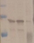
Zea mays crude extract (1), Zea mays mitochondria enriched fraction (2), immunoprecipitated sample (3), recombinant MHD4, expressed inE.coli (4). Total maize leaf proteins were extracted from 600 mg frozen leaf material with 1:4 ratio using 2.4 ml of extraction buffer (50 mM Trsi-HCl pH 8.0, 10 mM Ascorbate, 2 mM EDTA and 330 mM Mannitol, 1 x protease Inhibitor cocktail (Sigma Aldrich P8849-5ML). The homogenate was produced by mortar and pestle and centrifuged at 4000 x g for 10 min. at 4°C. The SDS-sample buffer (lane 1 in SDS-PAGE) is related to extraction using directly mortar and pestle and 2.4 ml of SDS-sampe buffer (0.1 M Tris-HCl pH 8.0, 100 mM DTT, 2 mM EDTA, 30% glycerol, 0.05 % bromophenolblue). The supernatant of 4000 x g was further centrifuged at 20,000 x g for 3 min in order to remove the chloroplasts. SDS-PAGE was performed using NuPAGE 4-12% (Invitrogen) at 120 V for 1.2 hours and blotted for 1h to Nitrocellulose membrane (BIOTrace NT, PALL, Life Sciences) using semi-dry transfer apparatus (Hoeffer) at 45 Ampere x 2 (gels) and variable voltage. Blots were blocked with Genscript 5M Quick Blocking solution (One step western blot kit, mix of solution A, casein hydrolysate and B, TTBS 1:1) for 15 minutes at room temperature (RT) with agitation. Blot was incubated in the primary antibody at a dilution of 1: 2 000 overnight at RT with agitation (sealed in a bag). The antibody solution was decanted and the blot was rinsed briefly twice with TTBS, then washed 3 times for 10 min in TTBS at RT with agitation. Blot was incubated in secondary antibody (anti-rabbit IgG horse radish peroxidase conjugated, diluted to 1:5000 in TTBS for 45 minutes at RT with agitation. The blot was washed as above and developed for 5 min with NBT/BCIP Ready-to-Use Tablets. Color development was stopped by decanting the solution and replacing with distilled water containing 10 mM EDTA. The membranes were dried on paper and photographed. Samples (boiled in boiling water for 10 minutes): 1) crude leaf extract by SDS-sample buffer (centrifuged 4000 x g for 10 min. at 4°C); Loaded 10µl 2) Leaf, mitochondria enriched supernatant 20,000 x g for 3 min. at 4°C; Loaded 10µl 3) Immunoprecipitation (IP) samples (tot. vol= 100µl); Loaded 8µl 4) Recombinant protein 1 µg/µl; loaded 1µl;
Courtesy of Dr. Giuseppe Dionisio, Aarhus University, Danmark
- Additional Information
-
Additional information (application): Immunoprecipitation was performed by using Dynabeads ® Protein A: briefly 100 µl suspension was washed with 200 µl TTBS (Tris Buffered saline, 50 mM Tris-HCl pH 7.6 and 165 mM NaCl with 0.1% Tween 80) using the magnetic stands for concentrating the magnetic beads. After wash the beads were preincubated with 20 µl primary antibodies in 180 µl TTBS at room temperature for 30 minutes (minimum 15 minues). A first wash was followed afterwards with 200 µl TTBS and hence a real incubation with 200 µl plant extract (supernatant 20,000 x g for 3 min.), 200µl of TTBS and further 50 µl YeastBuster reagent (Novagen) containing a mixture of detergents to break and solubilize the mitochondria membrane. This incubation at room temperature was allowed to be under mild shaking to allow the beads to be in suspension. Hence supernatant was aspirated away by the use of the magnetic stand and two further washesing steps with 200 µl TTBS were performed prior mixing with 100 µl SDS-Sample buffer.
Antibody is recognizing recombinant PGM1, overexpressed in E.coli. - Background
-
Background: Phosphoglucomutase is an enzyme which participates in both the breakdown and synthesis of glucose.
- Reviews:
-
This product doesn't have any reviews.


