1

Anti-JAR1 | Jasmonic acid-amido synthetase JAR1
AS16 3688 | Clonality: Polyclonal | Host: Rabbit | Reactivity: Arabidopsis thaliana
- Product Info
-
Immunogen: KLH-conjugated synthetic peptide derived from Arabidopsis thaliana JAR1 protein sequence, UniProt: Q9SKE2, TAIR: At2g46370 Host: Rabbit Clonality: Polyclonal Purity: Immunogen affinity purified serum in PBS pH 7.4. Format: Lyophilized Quantity: 50 µg Reconstitution: for reconstitution add 50 µl, of sterile water Storage: Store lyophilized/reconstituted at -20°C; once reconstituted make aliquots to avoid repeated freeze-thaw cycles. Please remember to spin the tubes briefly prior to opening them to avoid any losses that might occur from material adhering to the cap or sides of the tube. Tested applications: Western blot (WB) Recommended dilution: 1: 1000 (WB) Expected | apparent MW: 64 kDa (Arabidopsis thaliana) - Reactivity
-
Confirmed reactivity: Arabidopsis thaliana
Predicted reactivity: Capsicum annuum, Gossypium hirsutum, Nicotiana tabacum, Oryza sativa, Prunus yedoensis var. nudiflora, Solanum tuberosum, Ulmus americana
Species of your interest not listed? Contact usNot reactive in: - Application Examples
-
Application example 
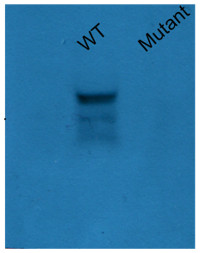
5 µg of protein from Arabidopsis thaliana Col0 and jar1-1 mutant were extracted using basic phenol protocol. 1g were grinded with liquid nitrogen with mortair and pestle, powder were resuspended in 3 mL of protein buffer (0.5 M Tris-HCl, 0.7 M sucrose, 1 mM PMSF, 50 mM EDTA, 0.1 M KCl and 0.2% β-mercaptoethanol; pH 8.0). Homogenate were centifuged at 8000 rpm and supernatant were mixed with three volumes of basic phenol, the mixture were incubated with agitation at room temperature during 10 mins. The mixture were centrifuged at 10000 rpm at 4ºC during 30 min, phenolic phase were recuperated in new tube and were mixed with one volume of ammonium acetate 0.1M in methanol, the mixture were incubated at -20 ºC during 4 hours. The mixture were centrifuged at 10000 rpm at 4º C during 20 minutes and protein pellet were washed three times with ammonium acetate 0.1M in methanol. Finally proteins were resuspended in tris-HCl (50 mM, pH 8.0) and the protein concentration were determined according Bradford method. Samples of 5 and 10µg were separated on 15 % SDS-PAGE using tank (BioRad system) to transfer to nitrocellulose membrane during 1 hour at 400 A. Blots were blocked with skimmed milk (10%) for 1h at room temperature (RT) with agitation. Blot was incubated in the primary antibody at a dilution of 1: 1000 for 1h at RT with agitation. The antibody solution was decanted and the blot was rinsed briefly twice, then washed once for 15 min and 3 times for 5 min in TBS-T at RT with agitation. Blot was incubated in secondary antibody (anti-rabbit IgG horse radish peroxidase conjugated, from Agrisera AS09 602) diluted to 1:20 000 in for 1h at RT with agitation. The blot was washed as above and developed for 5 min with ECL according to the manufacturer's instructions. Exposure time was 30 and 60 seconds. For transference control the membrane was stained with Ponceau red and integrity of proteins was evaluated using 12% SDS-PAGE comassie blue stained.
Courtesy of Dr. Rodrigo Contreras, Universidad de Santiago de Chile, ChileApplication examples: 
Reactant: Lupinus luteus (European yellow lupine)
Application: Western Blotting
Pudmed ID: 35214860
Journal: Plants (Basel)
Figure Number: 5A
Published Date: 2022-02-15
First Author: Domagalski, K.
Impact Factor:
Open PublicationJASMONATE RESISTANT1 (JAR1) is induced in the abscission zone (AZ) cells of yellow lupine flowers in response to soil drought. AZ from flowers collected from 48 day-old plants growing under optimal soil moisture (70% water holding capacity, WHC) was a control, while the “drought” variant was AZ excised from 48 day-old plants subjected to drought (25% WHC), as written precisely in the Material and Methods section. Western blot analysis was performed with a JAR1 antibody and revealed the presence of a reactive band of ~64 kDa (A). The membrane was scanned, densitometry analysis was performed (100% was set for the control), and presented in (B). Protein samples separated by SDS-PAGE and stained with Coomassie Brilliant Blue (C). Protein marker (M) is displayed on the left and sizes in kDa are provided. Immunolocalization of JAR1 in flower AZ cells during soil water deficit (E) and under control conditions (D). Green fluorescence indicates JAR1 localization (marked by red arrows). Blue labeling corresponds to DAPI-stained nuclei. AZ region is restricted by white dotted lines. Abbreviation: VB—vascular bundle. Bars = 40 µM. A significant difference is ^^p < 0.01.

Reactant: Lupinus luteus (European yellow lupine)
Application: Western Blotting
Pudmed ID: 35214860
Journal: Plants (Basel)
Figure Number: 5C
Published Date: 2022-02-15
First Author: Domagalski, K.
Impact Factor:
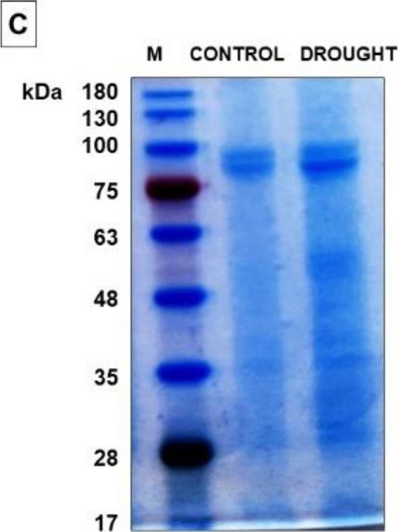
Open PublicationJASMONATE RESISTANT1 (JAR1) is induced in the abscission zone (AZ) cells of yellow lupine flowers in response to soil drought. AZ from flowers collected from 48 day-old plants growing under optimal soil moisture (70% water holding capacity, WHC) was a control, while the “drought” variant was AZ excised from 48 day-old plants subjected to drought (25% WHC), as written precisely in the Material and Methods section. Western blot analysis was performed with a JAR1 antibody and revealed the presence of a reactive band of ~64 kDa (A). The membrane was scanned, densitometry analysis was performed (100% was set for the control), and presented in (B). Protein samples separated by SDS-PAGE and stained with Coomassie Brilliant Blue (C). Protein marker (M) is displayed on the left and sizes in kDa are provided. Immunolocalization of JAR1 in flower AZ cells during soil water deficit (E) and under control conditions (D). Green fluorescence indicates JAR1 localization (marked by red arrows). Blue labeling corresponds to DAPI-stained nuclei. AZ region is restricted by white dotted lines. Abbreviation: VB—vascular bundle. Bars = 40 µM. A significant difference is ^^p < 0.01.

Reactant: Lupinus luteus (European yellow lupine)
Application: Immunocytochemistry
Pudmed ID: 35214860
Journal: Plants (Basel)
Figure Number: 5D,E
Published Date: 2022-02-15
First Author: Domagalski, K.
Impact Factor:
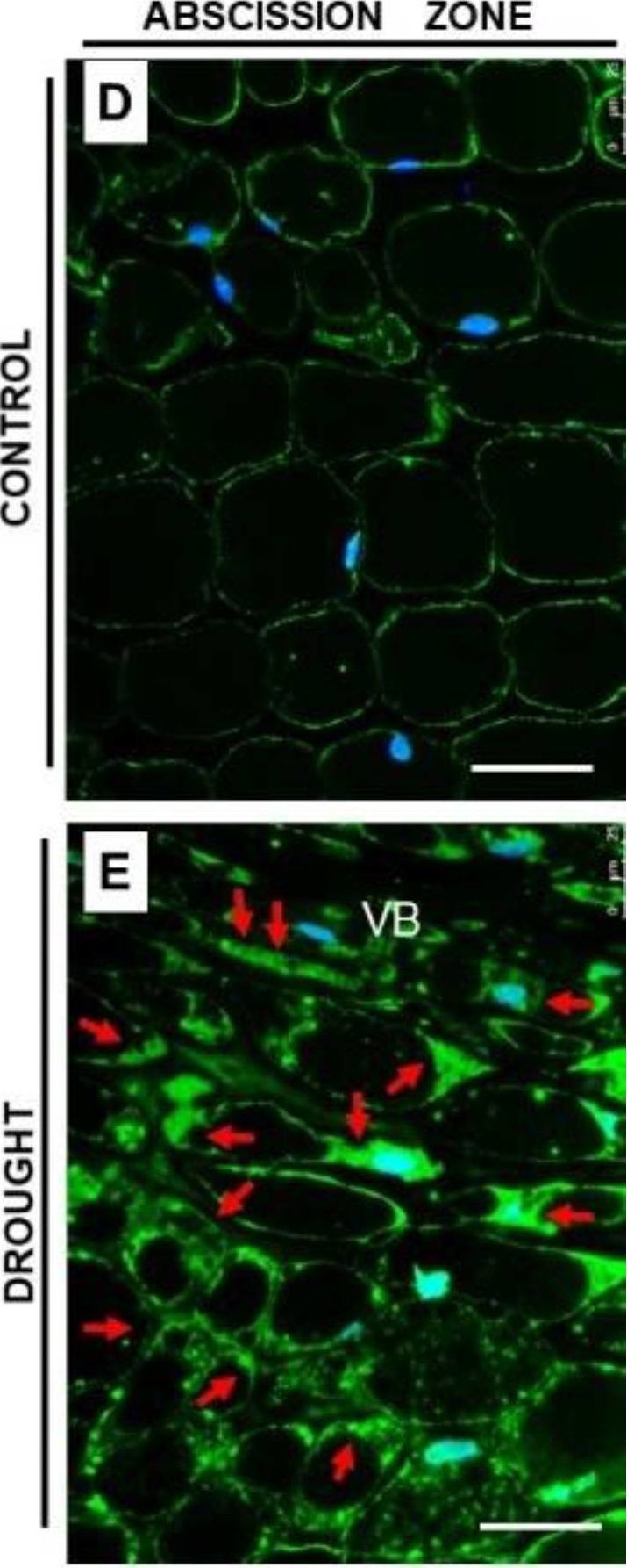
Open PublicationJASMONATE RESISTANT1 (JAR1) is induced in the abscission zone (AZ) cells of yellow lupine flowers in response to soil drought. AZ from flowers collected from 48 day-old plants growing under optimal soil moisture (70% water holding capacity, WHC) was a control, while the “drought” variant was AZ excised from 48 day-old plants subjected to drought (25% WHC), as written precisely in the Material and Methods section. Western blot analysis was performed with a JAR1 antibody and revealed the presence of a reactive band of ~64 kDa (A). The membrane was scanned, densitometry analysis was performed (100% was set for the control), and presented in (B). Protein samples separated by SDS-PAGE and stained with Coomassie Brilliant Blue (C). Protein marker (M) is displayed on the left and sizes in kDa are provided. Immunolocalization of JAR1 in flower AZ cells during soil water deficit (E) and under control conditions (D). Green fluorescence indicates JAR1 localization (marked by red arrows). Blue labeling corresponds to DAPI-stained nuclei. AZ region is restricted by white dotted lines. Abbreviation: VB—vascular bundle. Bars = 40 µM. A significant difference is ^^p < 0.01.
- Additional Information
-
Additional information (application): Phenol protein extraction method protocol. - Background
-
Background: JAR1 (Jasmonic acid-amido synthetase JAR1; jasmonate resistant 1) is catalyzing the synthesis of jasmonates-amino acid conjugates by adenylation. Plays an important role in the accumulation of JA-Ile in response to wounding, both locally and systemically; promotes JA responding genes especially in distal part of wounded plants, via the JA-Ile-stimulated degradation of JAZ repressor proteins by the SCF(COI)E3 ubiquitin-protein ligase pathway. Involved in the apoptosis-like programmed cell death (PCD) induced by fungal toxin fumonisin B1-mediated (FB1). Contributes to the sensitivity toward F.graminearum.
Alternative names: Jasmonate-amino acid synthetase JAR1, Protein FAR-RED INSENSITIVE 219, Protein JASMONATE RESISTANT 1 - Protocols
-
Phenol protein extraction protocol
- One gram of fresh leaf tissue was grinded in a mortar using liquid nitrogen to form a fine powder.
- The powder was resuspended in 3 mL of high density extraction buffer (0.5 M Tris-HCl, 0.7 M sucrose, 1 mM PMSF, 50 mM EDTA, 0.1 M KCl and 0.2% β-mercaptoethanol; pH 8.0).
- The homogenate was transferred to a conic tube and was mixed in an orbital shaker on the ice during 10 min. 1 mL of saturated phenol on Tris-HCl (pH 6.6-8.0) were added and the homogenate was mixed in an orbital shaker on the ice during 10 min.
- The mixture was centrifuged at 4 ºC during 10 min at 5000 ´g, the organic phase was recovered on a new tube and 4 volumes of ammonium acetate 0.1 M on methanol were added, the solution was mixed in a vortex and was stored at -20ºC overnight.
- The mixture was centrifuged at 4500xg for 15 min at 4ºC and pellet was washed twice with ammonium acetate 0.1 M on methanol and one with acetone, the pellet was dried at room temperature.
- Proteins were resuspended to saturation in Tris-HCl (50 mM, pH 8.0) and quantified according Bradford methodology (Bradford, 1976).
Courtesy of Dr. Rodrigo Contreras, Universidad de Santiago de Chile, ChileAgrisera Western Blot protocol and video tutorials
Protocols to work with plant and algal protein extracts
Agrisera Educational Posters Collection - Reviews:
-
This product doesn't have any reviews.


