1
Anti-HY5 | Protein long hypocotyl 5
AS12 1867 | Clonality: Polyclonal | Host: Rabbit | Reactivity: Arabidopsis thaliana
- Data sheet
-
- Product Info
-
Immunogen: KLH-conjugated peptide, derived from Arabidopsis thaliana HY5 protein sequence, UniProt:O24646 , TAIR: AT5G11260. Chosen peptide is not conserved in HY5 protein sequence. Host: Rabbit Clonality: Polyclonal Purity: Immunogen affinity purified serum in PBS pH 7.4. Format: Lyophilized Quantity: 50 µg Reconstitution: For reconstitution add 50 µl of sterile water Storage: Store lyophilized/reconstituted at -20°C; once reconstituted make aliquots to avoid repeated freeze-thaw cycles,Please remember to spin the tubes briefly prior to opening them to avoid any losses that might occur from material adhering to the cap or sides of the tube. Tested applications: Western blot (WB) Recommended dilution: 1: 500 to 1 : 1000 (WB) Expected | apparent MW: 18.5 kDa - Reactivity
-
Confirmed reactivity: Arabidopsis thaliana Predicted reactivity: Brassica pekinensis
Species of your interest not listed? Contact usNot reactive in: Citrus reticulata, Hordeum vulgare, Marchantia polymorhpa, Oryza sativa, Pisum sativum, Populus sp., Solanum lycopersicum, Triticum aestivum, Zea mays
- Application Examples
-
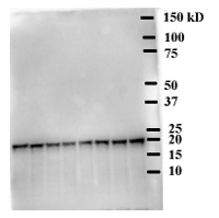
80 μg/well of total protein extracted freshly from the shoot part of 50 Arabidopsis thaliana seedlings, grown for 7 days at white light. Extraction buffer composition was 100 mM Tris pH 7.5, 1 mM EDTA, 8 M urea, 1× protease inhibitor cocktail, 2 mM PMSF. The extract was cleared by centrifugation at 16000g for 10 min at 4°C. 6× SDS sample buffer was added and the boiled for 10 min, centrifuged for 2 min and then separated on a 10% SDS-PAGE. The gel was blotted onto polyvinylidene difluoride membranes (0.2 µm pore) using wet transfer in the cold. Blot was blocked with 5 % nonfat milk for 1h at RT with agitation. Blot was incubated in the primary antibody (anti-HY5, AS12 1867) at a dilution of 1: 10 000 overnight with agitation in TBST with 1% BSA. The antibody solution was decanted, and the blot was washed three times for 10 minutes with TBS-T at RT with agitation. Blot was incubated in matching secondary antibody (anti-rabbit IgG horse radish peroxidase conjugated) diluted to 1: 10 000 in for 2h at RT with agitation. The blot was washed as above and then developed with the following chemiluminescent detection reagent: ECL kit. Exposure time was 1 min 40 sec.Courtesy od prof. Enamul Huq, University of Texas at Austin, USA

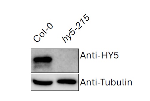
10 µg of total protein extracted freshly from 7-d old Arabidopsis thaliana seedlings using Trichloroacetic acid and Acetone (Mechin et al. 2007), and denatured with LDS (Lithium dodecyl sulfate) sample buffer at 70°C for 10 min. Proteins were separated on 12% SDS-PAGE and blotted 7 min to PVDF (pore size of 0.2 um), using semi-dry transfer. Blot was blocked with 5% milk for 4°C/ON with agitation. Blot was incubated in the primary antibody at a dilution of 1: 1 000 for 2 h/RT with agitation in TBS-T. The antibody solution was decanted and the blot was rinsed briefly, then washed three times for 15 min in TBS-T at RT with agitation. Blot was incubated in Agrisera matching secondary antibody (anti-rabbit IgG horse radish peroxidase conjugated, AS09 602) diluted to 1:10,000 in for 1 h/RT with agitation. The blot was washed as above and developed for 2 min with chemiluminescence detection reagent. Exposure time was 100 seconds.
The seedlings were grown 4 d in dark and 3 d in continuous light (~120 uE). Seedlings were ground in whole for protein extraction.
Courtesy of Xin Hou, Pogson Lab, Research School of Biology, ANU College of Science, Australia
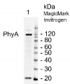
20 ug of total protein from control (1), 35S::YFP-HY5-HA (2, red arrow), 35S::YFP-HY5-HA + 35S::CFP-X protein (green arrow), were separated on 12 % SDS-PAGE using tank transfer and blotted 1 h to PVDF (Biorad). Blots were blocked with 5 % skim milk for 1h at room temperature (RT) with agitation. Blot was incubated in the anti-HY5 antibody (second panel from the left) at a dilution of 1: 1000 for 1h at RT with agitation. The antibody solution was decanted and the blot was rinsed briefly twice, then washed once for 15 min and 3 times for 5 min. in PBS-T at RT with agitation. Blot was incubated in secondary antibody (anti-rabbit IgG, HRP conjugated from Agrisera, AS09 602), diluted to 1: 10 000 for 1h at RT with agitation. The blot was washed as above and developed for 5 min. with chemiluminescent detection, according to the manufacturer's instructions. Exposure time was 60 seconds.
Courtesy of Dr. Seok Keun Cho, University of Copenhagen, DanmarkApplication examples: 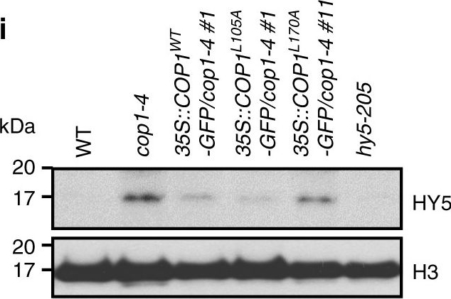
Reactant: Arabidopsis thaliana (Thale cress)
Application: Western Blotting
Pudmed ID: 29273730
Journal: Nat Commun
Figure Number: 5I
Published Date: 2017-12-22
First Author: Lee, B. D., Kim, M. R., et al.
Impact Factor: 13.783
Open PublicationCOP1 variants that are unable to homo-dimerize are non-functional. a Mutation sites in the CLS motif of COP1. b COP1L170A forms dimers poorly when compared with COP1WT or COP1L105A in N. benthamiana. c FKF1 interacts more weakly with COP1L170A (poor dimer formation) than COP1L105A (normal dimer formation) in N. benthamiana. d Various transgenic plants at bolting under LD and SD. Scale bars, 2?cm. e Flowering times of the plants in d. The number of rosette leaves at bolting represents the flowering time of each genotype. Data are means?±?s.d. of 20 plants. f CO accumulation of each genotype in d. Ten-day-old plants were harvested at ZT15 (1?h before dark) in LD. g Hypocotyl elongation of plants in d grown for 5 days in complete darkness. Scale bars, 1?cm. h Hypocotyl length of transgenic plants in darkness. Data are means?±?s.d. of 20 plants. i HY5 accumulation in various transgenic plants
Reactant: Arabidopsis thaliana (Thale cress)
Application: Western Blotting
Pudmed ID: 32003746
Journal: Elife
Figure Number: 7D
Published Date: 2020-01-31
First Author: Cazzonelli, C. I., Hou, X., et al.
Impact Factor: 7.448
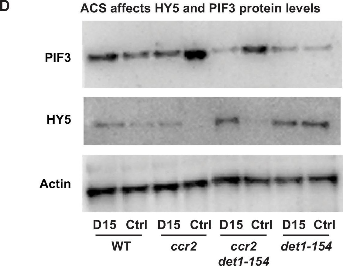
Open PublicationChemical inhibition of CCD activity revealed how a ccr2 generated apocarotenoid signal transcriptionally up-regulates POR and PIF3 in parallel to det1-154 during skotomorphogenesis.(A) Transcript levels of PORA, PIF3 and HY5 in WT, ccr2, ccr2 det1-154 and det1-154 etiolated seedlings growing on MS media (+ /- D15). Statistical analysis denoted as a star was performed by a pair-wise t-test (p<0.05). Error bars represent standard error of means. (B), (C) and (D) Representative western blot images showing POR, DET1, PIF3 and HY5 protein levels, respectively. Proteins were extracted from WT, ccr2 and ccr2 det1-154 etiolated seedlings grown on MS media without (control; Ctrl) or with the chemical inhibitor of CCD activity (D15). The membrane was re-probed using anti-Actin antibody as an internal loading control. Lattice-like symbol below POR western (B), represents formation of a PLB in etiolated cotyledons from that genotype and treatment. (E) Model describing how a cis-carotene derived cleavage product, ACS, regulates POR, HY5, PIF3 and PLB formation during skotomorphogenesis. DET1 maintains skotomorphogenesis by post-transcriptionally maintaining a higher and lower PIF3 and HY5 protein levels, respectively. HY5 promotes and PIF3 represses PhANG expression. det1 mutants trigger photomorphogenesis in that they lack POR mRNA transcripts, protein and a PLB. ccr2 generates ACS that enhances POR mRNA transcript and protein levels that enable PLB formation in det1-154. det1-154 restores PLB formation in ccr2 by blocking a signalling pathway acting independent of POR.The DET1-154 peptide is smaller in det1-154 mutant genotypes.(A) Representative western blot image showing the reduced DET1 peptide size in ccr2 det1-154 and det1-154 (59 kDa) compared to WT and ccr2 (62 kDa). Gel electrophoresis of the gel membrane from Figure 7C (electrophoresis for 38 min at 165 volt) was extended for 120 min at 100 volts to resolve the 3 kDa difference in DET1-154 protein size. Under these conditions, the 37 kDa ACTIN peptide and pre-stained ladder were not detected on the membrane. Proteins were extracted from WT, ccr2 and ccr2 det1-154 etiolated seedlings grown on MS media without (control; Ctrl) or with the chemical inhibitor of CCD activity (D15).
Reactant: Arabidopsis thaliana (Thale cress)
Application: Western Blotting
Pudmed ID: 32003746
Journal: Elife
Figure Number: 8B
Published Date: 2020-01-31
First Author: Cazzonelli, C. I., Hou, X., et al.
Impact Factor: 7.448
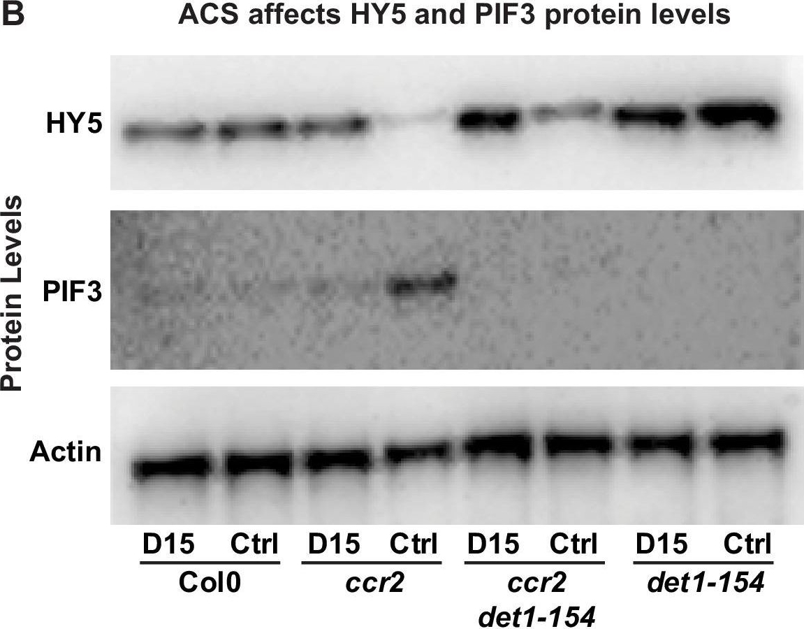
Open PublicationChemical inhibition of CCD activity revealed how a ccr2 generated apocarotenoid signal transcriptionally represses HY5 and LHCB2 expression during photomorphogenesis.(A) Transcript levels of PIF3 and HY5 in WT, ccr2, ccr2 det1-154 and det1-154 de-etiolated seedlings growing on MS media + /- D15. (B) Representative western blot images showing PIF3 and HY5 protein levels in WT, ccr2, ccr2 det1-154 and det1-154 de-etiolated seedlings growing on MS media + /- D15. The membrane was re-probed using anti-Actin antibody as an internal loading control. (C) Protein and transcript levels of LHCB2 expression in WT and ccr2 de-etiolated seedlings growing on MS media + /- D15. (D) Model showing how ACS regulates HY5 and LHCB2 expression in ccr2. Images of seedlings represent are cotyledons are coloured green or yellow to reflect the delay in chlorophyll biosynthesis induced by ACS as evidenced in Figure 6c. De-etiolation of seedlings was performed by transferring 4-d-old etiolated seedlings to continuous light for 3 d to induce photomorphogenesis. Statistical analysis denoted as a star was performed by pair-wise t-test (p<0.05). Error bars represent standard error of means. Ctrl; Control; Ctrl, D15; chemical inhibitor of CCD activity.
- Additional Information
-
Additional information (application): This antibody detects recombinant HY5 in Nicotiana benthamiana.
Extraction and loading buffer with 6-8 M urea buffer needs to be used when working with endogenous extract to allow detection with this antibody or TCA/acetone protein extraction or as described in Mechin et al. (2007).
Samples need to be harvested under dim-green safe light conditions, to avoid degradation during harvesting and extraction process. - Background
-
Background: HY5 (Protein long hypocotyl 5) is a light-regulated transcription factor that promotes photomorphogenesis in light. Involved in the blue light specific pathway. Heterodimer. Expressed in root, hypocotyl, cotyledon, leaf, stem and floral organs. Localized in nucleus. Alternative names: Protein LONG HYPOCOTYL 5, bZIP transcription factor 56, AtbZIP56. - Product Citations
-
Selected references: Malakar et al. (2025). BBX24/BBX25 antagonizes the function of thermosensor ELF3 to promote PIF4-mediated thermomorphogenesis in Arabidopsis. Plant Commun. 2025 May 28:101391.doi: 10.1016/j.xplc.2025.101391.
Yao et. al. 2024).Cooperative transcriptional regulation by ATAF1 and HY5 promotes light-induced cotyledon opening in Arabidopsis thaliana. Sci Signal. 2024 Jan 2;17(817):eadf7318.
Liu et al. (2024). Phosphorylation of Arabidopsis UVR8 photoreceptor modulates protein interactions and responses to UV-B radiation. Nat Commun. 2024 Feb 9;15(1):1221.doi: 10.1038/s41467-024-45575-7.
Cazzonelli et al. (2019). A cis-carotene derived apocarotenoid regulates etioplast and chloroplast development. https://doi.org/10.1101/528331
Lee et al. (2017). The F-box protein FKF1 inhibits dimerization of COP1 in the control of photoperiodic flowering. Nat Commun. 2017 Dec 22;8(1):2259. doi: 10.1038/s41467-017-02476-2.
Sinclair et al. (2017) Etiolated Seedling Development Requires Repression of Photomorphogenesis by a Small Cell-Wall-Derived Dark Signal. Curr Biol. 2017 Nov 20;27(22):3403-3418.e7. doi: 10.1016/j.cub.2017.09.063. - Protocols
-
Agrisera Western Blot protocol and video tutorials
Protocols to work with plant and algal protein extracts - Reviews:
-
A. R. | 2021-11-11The antibody worked as expected for Arabidopsis thaliana protein extracts. The antibody can be recommended.

