1

Anti-H3 | Histone H3 (chicken antibody)
AS15 2855 | Clonality: Polyclonal | Host: Chicken | Reactivity: A. thaliana, C. reinhardtii, L. esculentum, T. aestivum | cellular [compartment marker] of nucleoplasm
- Product Info
-
Immunogen: KLH-conjugated synthetic peptide derived from known H3 sequences, inluding Arabidopsis thaliana H3.3 P59169 (At4g40030,At4g40040,At5g10980), H3.2 P59226(At1g09200,At3g27360,At5g10390,At5g10400,At5g65360), H3-like 2 Q9FXI7 (At1g19890)
Host: Chicken Clonality: Polyclonal Purity: Purified, total IgY (chicken egg yolk immunoglobulin) in PBS pH 8. Contains 0.02 % sodium azide. Format: Liquid Quantity: 50 µl Storage: Store at 4°C. Please remember to spin the tubes briefly prior to opening them to avoid any losses that might occur from material adhering to the cap or sides of the tube. Tested applications: Western blot (WB) Recommended dilution: 1 : 2500 (WB) Expected | apparent MW: 15 | 17 kDa
- Reactivity
-
Confirmed reactivity: Arabidopsis thaliana, Chlamydomonas reinhardtii, Lycopersicum esculentum, Triticum aestivum Predicted reactivity: Arabidopsis thaliana, Brassica napus, Brassica oleracea, Capsicum annuum, Chlamydomonas acidophila, Galdieria sulphuraria, Hordeum vulgare, Medicago sativa, Nannochloropsis gaditana, Nicotiana tabacum, Oryza sativa, Physcomitrium patens, Pinus pinaster, Pisum sativum, Salicornia europaea, Solanum lycopersicum, Solanum sogarandinum, Solanum tuberosum, Triticum aestivum, Zea mays, Vicia faba, Vitis vinifera, Volvox sp.
Species of your interest not listed? Contact usNot reactive in: No confirmed exceptions from predicted reactivity are currently known - Application Examples
-
Application example 
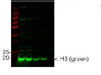
From left to right: 2 wells of nuclear + cytoplasmic enriched protein, 2 wells of total proteins of Chlamydomonas reinhardtii protein saturated in 8M urea were separated on 15% SDS-PAGE and blotted for 1hour to 0.2 µm nitrocellulose at 100V using wet transfer system. Blots were blocked with 0.5% cold fish gelatin for 1hr at room temp with agitation. Blot was incubated in the primary antibody (anti-H3) at a dilution of 1:2500 for an hour at RT with agitation. The blots were washed with 3X 15min TBS-TT at RT with agitation. Blots as incubated in the secondary antibody (AS11 1827, Goat anti-Chicken IgY (H&L), DyLight® 800 conjugated, Agrisera) 1:5000 dilution for 30min at RT with agitation and washed 1X with TBSTT for 15min, 1X with TBST for 15min before scanning with the ODyssey IRD scanner.
Courtesy of Dr. Betty Chung, University of Cambridge, United KingdomApplication examples: 
Reactant: Chlamydomonas reinhardtii (Green Alga)
Application: Western Blotting
Pudmed ID: 31366981
Journal: Sci Rep
Figure Number: 2C
Published Date: 2019-07-31
First Author: Chung, B. Y. W., Valli, A., et al.
Impact Factor: 4.13
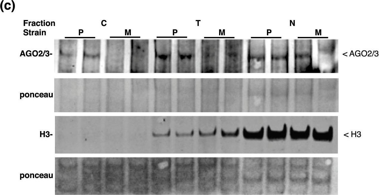
Open PublicationExpression and differential localisation of AGO2 and AGO3. (a) Antigen sites utilised for peptide-based antibody production. (b) Western blots for total and ?-2/3-PAZ-immunoprecipitated product using extracts from either the parental (P) line or ago3-25 (M). At-AGO1 antibody was utilized as an IP negative control. (c) Differential localisation of Cr-AGO2 and 3. Total protein isolated from cytoplasmic, total and nuclear fractions are indicated as C, T and N, respectively. Extracts from either Parental or ago3-25 strain are labelled P and M, respectively. Protein bands corresponding to AGO2/3 or Histone 3 (H3) were indicated.
- Additional Information
-
Additional information: Cellular [compartment marker] of nucleoplasm, loading control antibody for Chlamydomonas reinhardtii - Background
-
Background: Histone 3 (H3) located in nuclei, incorporated into chromatin. Present in nucleosome together with H2A, H2B and H4.
- Product Citations
-
Selected references: Loudya et al. (2021) Cellular and transcriptomic analyses reveal two-staged chloroplast biogenesis underpinning photosynthesis build-up in the wheat leaf. Genome Biol. 2021 May 11;22(1):151. doi: 10.1186/s13059-021-02366-3. PMID: 33975629; PMCID: PMC8111775.
Chung et al. (2019) Distinct roles of Argonaute in the green alga Chlamydomonas reveal evolutionary conserved mode of miRNA-mediated gene expression. Sci Rep. 2019 Jul 31;9(1):11091. doi: 10.1038/s41598-019-47415-x. - Protocols
-
Agrisera Western Blot protocol and video tutorials
Preparation of cytosolic and nuclear protein fractions
1. Prepare protoplasts from 50 ml Arabidopsis thaliana cell culture according to the protocol of PEG transfection.
2. Resuspend protoplasts in 10 ml GH buffer and keep the solution on ice for 10 min.
GH buffer: 100mM glycine
0.1% Hexylene glycol
0.37M (4.7% w/v) saccharose
0.3mM Spermine
1.0mM Spermidine
pH 8.3 with Ca(OH)2
3. To release nuclei add Triton X100 to a final concentration of 0.1%. Pipetting gently up and down several
times with a plastic pipette might be necessary to lyse cells.
4. After 5min sediment nuclei by centrifugation at 1000 xg for 15 min at 4°C. Save supernatant as the
cytoplasmic fraction. Wash the pelleted nuclei two times with GHT (GH+0.1% TX100) then finally
resuspended in a suitable volume of extraction buffer + protease inhibitors.Courtesy Dr. Laszlo Bako, Umeå Plant Science Centre, Sweden
- Reviews:
-
This product doesn't have any reviews.


