1

Anti-COI1 | Coronate insensitive 1 (rabbit antibody)
AS12 2637 | Clonality: Polyclonal | Host: Rabbit | Reactivity: Arabidopsis thaliana
- Product Info
-
Immunogen: KLH-conjugated synthetic peptide derived from Arabidopsis thaliana COI1 protein, UniProt: O04197 , TAIR: At2g39940
Host: Rabbit Clonality: Polyclonal Purity: Immunogen affinity purified serum in PBS pH 7.4. Format: Lyophilized Quantity: 50 µg Reconstitution: For reconstitution add 25 µl of sterile water Storage: Store lyophilized/reconstituted at -20°C; once reconstituted make aliquots to avoid repeated freeze-thaw cycles. Please remember to spin the tubes briefly prior to opening them to avoid any losses that might occur from material adhering to the cap or sides of the tube. Tested applications: Western blot (WB) Recommended dilution: 1 : 1000 (WB) Expected | apparent MW: 70 kDa
- Reactivity
-
Confirmed reactivity: Arabidopsis thaliana Predicted reactivity: Brassica rapa, Glycine max, Lupinus luteus, Medicago tribuloides, Nicotiana tabacum, Oryza sativa, Pisum sativum, Populus trichocarpa, Ricinus communis, Solanum lycopersicum, Vitis vinifera
Species of your interest not listed? Contact usNot reactive in: No confirmed exceptions from predicted reactivity are currently known - Application Examples
-

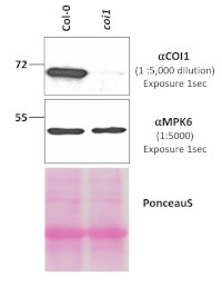
Total proteins were isolated from 7-day old Arabidopsis thaliana Col-0 (wild-type) and coi1 (null mutant) seedlings according to [ref 1]. Proteins were denatured at 65°C for 5 min. 30 μg of proteins were separated on a 10% SDS-PAGE and transferred for 70 min at 55V using a tank transfer system to nitrocellulose membrane. Blots were blocked with 1x PBS + 0.1% Tween 20 (PBS- T) + 5% milk for 1hr at room temperature (RT) with agitation. Blots were incubated in primary ⍺-COI1 antibody (AS12 2637) diluted to 1:5,000 in PBS-T+5 % milk at 4°C overnight with agitation. The primary antibody solution was decanted, and the blots were washed four times (6-8 minutes each) in 1x PBS-T at RT with agitation prior to incubation with secondary antibody Goat anti Rabbit IgG (H&L) – HRP conjugated (AS09 602-trial) diluted to 1:20,000 in 1x PBS-T + 5% milk for 2hr at RT with agitation. The blots were washed as above and developed for 4 min with chemiluminescent detection reagent ECL Bright (AS16 ECL-N). Exposure times (in seconds) to X-ray films were done as indicated. PonceauS stained membrane and membrane portion probed with ⍺-MPK6 served as loading controls [ref 1].
Courtesy of Alani Antoine-Mitchell and Antje Heese, University of Missouri, Div. Biochemistry, IPG (USA)
References: [1] LaMontagne, ED., Collins, CA., Peck, S.C., and Heese, A. 2016. Isolation of microsomal membrane proteins from Arabidopsis thaliana. Curr. Protoc. Plant Biol. 1:217-234. doi: 10.1002/cppb.20020Application examples: 
Reactant: Lupinus luteus (European yellow lupine)
Application: Immunocytochemistry
Pudmed ID: 35214860
Journal: Plants (Basel)
Figure Number: 6D
Published Date: 2022-02-15
First Author: Domagalski, K.
Impact Factor:
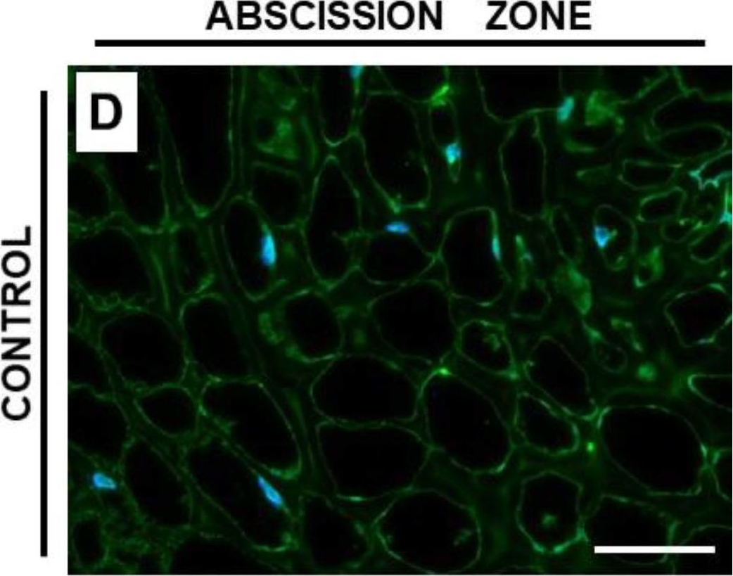
Open PublicationThe content of coronatine insensitive 1 (COI1) in the flower abscission zone (AZ) of Lupinus luteus strongly increased under soil water deficit. Flower AZ was harvested from plants growing in the soil of optimal moisture (70% water holding capacity, WHC, control) or drought-stressed lupines (25% WHC). Detailed description in the Material and Methods section. Proteins were isolated and subjected to Western blot analysis with an anti-COI1 antibody, which revealed the presence of a band of ~70 kDa (A). Quantitative densitometry analysis was made and presented in (B) (100% was set for the control). ^^p < 0.01 indicates a significant difference. Protein samples were separated by SDS-PAGE and stained with Coomassie Brilliant Blue (C). Mass marker (M) is provided on the left, sizes of proteins are displayed in kDa. COI1 antibody was used for immunolocalization analysis performed on control (D) and drought-stressed (E,F) tissues of AZ from flowers. A green fluorescence signal, indicated by red arrows, corresponds to the presence of COI1. Nuclei were detected by DAPI (blue color). Enlarged cells of stressed AZ are provided on the left side of the (F) image. These are magnified regions marked by white boxes and show colocalization of CO1 with nuclei (red arrowheads). Bars = 40 µM.

Reactant: Lupinus luteus (European yellow lupine)
Application: Immunocytochemistry
Pudmed ID: 35214860
Journal: Plants (Basel)
Figure Number: 6E,F
Published Date: 2022-02-15
First Author: Domagalski, K.
Impact Factor:
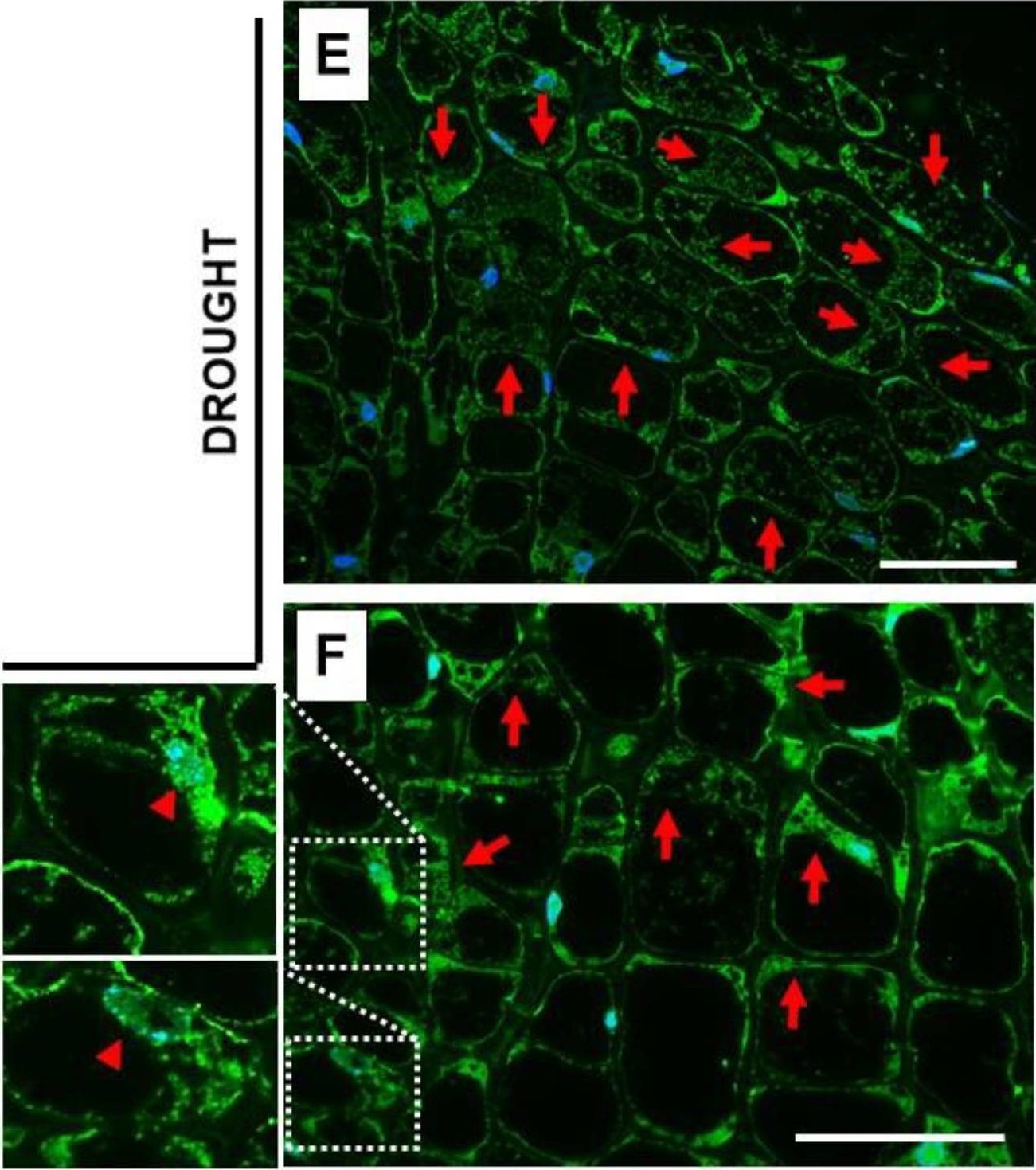
Open PublicationThe content of coronatine insensitive 1 (COI1) in the flower abscission zone (AZ) of Lupinus luteus strongly increased under soil water deficit. Flower AZ was harvested from plants growing in the soil of optimal moisture (70% water holding capacity, WHC, control) or drought-stressed lupines (25% WHC). Detailed description in the Material and Methods section. Proteins were isolated and subjected to Western blot analysis with an anti-COI1 antibody, which revealed the presence of a band of ~70 kDa (A). Quantitative densitometry analysis was made and presented in (B) (100% was set for the control). ^^p < 0.01 indicates a significant difference. Protein samples were separated by SDS-PAGE and stained with Coomassie Brilliant Blue (C). Mass marker (M) is provided on the left, sizes of proteins are displayed in kDa. COI1 antibody was used for immunolocalization analysis performed on control (D) and drought-stressed (E,F) tissues of AZ from flowers. A green fluorescence signal, indicated by red arrows, corresponds to the presence of COI1. Nuclei were detected by DAPI (blue color). Enlarged cells of stressed AZ are provided on the left side of the (F) image. These are magnified regions marked by white boxes and show colocalization of CO1 with nuclei (red arrowheads). Bars = 40 µM.

Reactant: Lupinus luteus (European yellow lupine)
Application: Western Blotting
Pudmed ID: 35214860
Journal: Plants (Basel)
Figure Number: 6A
Published Date: 2022-02-15
First Author: Domagalski, K.
Impact Factor:
Open PublicationThe content of coronatine insensitive 1 (COI1) in the flower abscission zone (AZ) of Lupinus luteus strongly increased under soil water deficit. Flower AZ was harvested from plants growing in the soil of optimal moisture (70% water holding capacity, WHC, control) or drought-stressed lupines (25% WHC). Detailed description in the Material and Methods section. Proteins were isolated and subjected to Western blot analysis with an anti-COI1 antibody, which revealed the presence of a band of ~70 kDa (A). Quantitative densitometry analysis was made and presented in (B) (100% was set for the control). ^^p < 0.01 indicates a significant difference. Protein samples were separated by SDS-PAGE and stained with Coomassie Brilliant Blue (C). Mass marker (M) is provided on the left, sizes of proteins are displayed in kDa. COI1 antibody was used for immunolocalization analysis performed on control (D) and drought-stressed (E,F) tissues of AZ from flowers. A green fluorescence signal, indicated by red arrows, corresponds to the presence of COI1. Nuclei were detected by DAPI (blue color). Enlarged cells of stressed AZ are provided on the left side of the (F) image. These are magnified regions marked by white boxes and show colocalization of CO1 with nuclei (red arrowheads). Bars = 40 µM.

Reactant: Lupinus luteus (European yellow lupine)
Application: Western Blotting
Pudmed ID: 35214860
Journal: Plants (Basel)
Figure Number: 6C
Published Date: 2022-02-15
First Author: Domagalski, K.
Impact Factor:
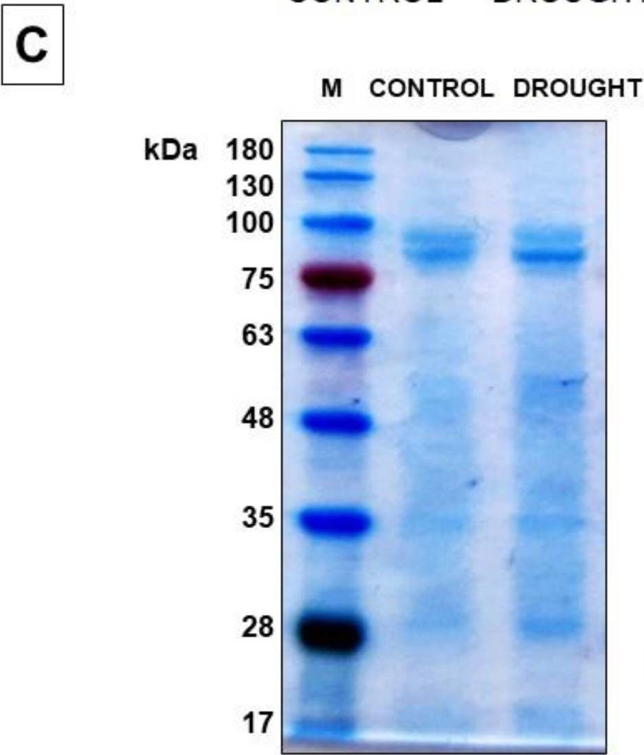
Open PublicationThe content of coronatine insensitive 1 (COI1) in the flower abscission zone (AZ) of Lupinus luteus strongly increased under soil water deficit. Flower AZ was harvested from plants growing in the soil of optimal moisture (70% water holding capacity, WHC, control) or drought-stressed lupines (25% WHC). Detailed description in the Material and Methods section. Proteins were isolated and subjected to Western blot analysis with an anti-COI1 antibody, which revealed the presence of a band of ~70 kDa (A). Quantitative densitometry analysis was made and presented in (B) (100% was set for the control). ^^p < 0.01 indicates a significant difference. Protein samples were separated by SDS-PAGE and stained with Coomassie Brilliant Blue (C). Mass marker (M) is provided on the left, sizes of proteins are displayed in kDa. COI1 antibody was used for immunolocalization analysis performed on control (D) and drought-stressed (E,F) tissues of AZ from flowers. A green fluorescence signal, indicated by red arrows, corresponds to the presence of COI1. Nuclei were detected by DAPI (blue color). Enlarged cells of stressed AZ are provided on the left side of the (F) image. These are magnified regions marked by white boxes and show colocalization of CO1 with nuclei (red arrowheads). Bars = 40 µM.
- Additional Information
-
Additional information (application): Antibody detects 100 ng of recombinant COI1 protein, expressed in Hi5 suspenstion insect cells.
The peptide used to elicit this antibody is partially conserved in Zea mays: ZmCOI1a, ZMCOI1b, ZMCO1c and ZmCOI2. - Background
-
Background: COI1 (Coronate insensitive 1) is an esential component of jasmonate receptor. Required for jasmonate-regulated plant fertility and defense processes, and for coronatine and/or other elicitors perceptions/responses. Alternative names: F-box/LRR-repeat protein 2, AtCOI1, AtFBL2. - Product Citations
-
Selected references: Agrawal et al. (2022) MEDIATOR SUBUNIT17 integrates jasmonate and auxin signaling pathways to regulate thermomorphogenesis. Plant Physiol. 2022 Aug 1;189(4):2259-2280. doi: 10.1093/plphys/kiac220. PMID: 35567489. - Protocols
-
Agrisera Western Blot protocol and video tutorials
Protocols to work with plant and algal protein extracts
Agrisera Educational Posters Collection
- Reviews:
-
Aashish Ranjan | 2021-09-30The antibody worked great for western blotting with Arabidopsis leaves. We have used 1:1000 dilution for the experiments and could clearly detect the differential protein levels across control and treatments.


