1

Anti-ATG9 | Autophagy-related protein 9 (C-terminal)
AS16 4072 | Clonality: Polyclonal | Host: Rabbit | Reactivity: Arabidopsis thaliana
- Product Info
-
Immunogen: KLH-conjugated peptide derived from Arabidopsis thaliana ATG9 protein sequence, UniProt: Q8RUS5, TAIR: AT2G31260
Host: Rabbit Clonality: Polyclonal Purity: Immunogen affinity purified serum in PBS pH 7.4. Format: Lyophilized Quantity: 50 µg Reconstitution: For reconstitution add 50 µl of sterile water Storage: Store lyophilized/reconstituted at -20°C; once reconstituted make aliquots to avoid repeated freeze-thaw cycles.Please remember to spin the tubes briefly prior to opening them to avoid any losses that might occur from material adhering to the cap or sides of the tube. Tested applications: Western blot (WB) Recommended dilution: 1 : 10 000 (WB)
Expected | apparent MW: 99.5 | 100 kDa - Reactivity
-
Confirmed reactivity: Arabidopsis thaliana Predicted reactivity: Aquilegia coerulea, Brassica napus, Capsella rubella, Daucus carota, Eutrema salsugineum, Gossypium hirsutum, Juglans regia, Lactuca sativa, Noccaea caerulescens, Theobroma cacao, Trema orientale Not reactive in: Nicotiana tabacum
- Application Examples
-
Application example

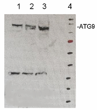
Increasing amount of microsomal fraction of Arabidopsis thaliana cell culture (1,2,3). Microsomal protein was extracted accoring to Maudoux et al. (2000). and denatured with loading buffer (2 % SDS) at 37°C for 1h before SDS-PAGE gel electrophoresis and blotted to PVDF using semi-dry. Blots were blocked with 3 % nonfat milk + 0.1 % goat serum in TBS-T (Tween 20) for 1h at room temperature (RT) with agitation. Blot was incubated in the primary antibody at a dilution of 1: 10 000 + 1 % goat serum for ON at 4°C with agitation. The antibody solution was decanted and the blot was rinsed briefly twice, then washed once for 15 min and 3 times for 5 min in TBS-T at RT with agitation. Blot was incubated in secondary antibody (anti-rabbit IgG horse radish peroxidase conjugated) diluted to 1:10 000 in TBS-T + 1 % goat serum for 1h at RT with agitation. The blot was washed as above and developed with chemiluminescent detection reagent. Exposure time was seconds.
Red marker band has apparent mass of 72 kDa.Courtesy of Dr. Henri Batoko, University of Louvain, Belgium
- Additional Information
-
Additional information (application): Plant ATG9 is most likely N-glycosylated, and glycosylated membrane proteins can migrate as more than one form in SDS-PAGE (smear); the protein most likely forms functional dimers which could be detected also after western blotting.
Usage of microsomal fraction is highly recommended. - Background
-
Background: ATG9 (Autophagy-related protein 9) is required for autophagy that plays an essential role in plant nutrient recycling.
Alternative name: AtAPG9. - Protocols
-
Agrisera Western Blot protocol and video tutorials
Protocols to work with plant and algal protein extracts
Oxygenic photosynthesis poster by prof. Govindjee and Dr. Shevela
Z-scheme of photosynthetic electron transport by prof. Govindjee and Dr. Björn and Dr. Shevela - Reviews:
-
This product doesn't have any reviews.


