1

Anti-AGO2 | Argonaute 2
AS13 2682 | Clonality: Polyclonal | Host: Rabbit | Reactivity: Arabidopsis thaliana
- Data sheet
- Product Info
-
Immunogen: KLH-conjugated synthetic peptide, derived from Arabidopsis thaliana AGO2 protein, UniProt: Q9SHF3, TAIR: AT1G31280 Host: Rabbit Clonality: Polyclonal Purity: Immunogen affinity purified IgG in PBS pH 7.4. Format: Lyophilized Quantity: 50 µg Reconstitution: For reconstitution add 50 µl of sterile water Storage: Store lyophilized/reconstituted at -20°C; once reconstituted make aliquots to avoid repeated freeze-thaw cycles. Please remember to spin the tubes briefly prior to opening them to avoid any losses that might occur from material adhering to the cap or sides of the tube. Tested applications: Immunoprecipitation (IP), Western blot (WB) Recommended dilution: 1 : 250-1 : 500 (WB) Expected | apparent MW: 113 kDa
- Reactivity
-
Confirmed reactivity: Arabidopsis thaliana
Predicted reactivity: Capsella rubella, Solanum tuberosum
Species of your interest not listed? Contact usNot reactive in: Brassica napus, Chlamydomonas reinhardtii, Medicago truncatula, Nicotiana benthamiana, Populus sp., Solanum lycopersicum, Zea mays
- Application Examples
-

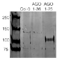
300 µg/well of Arabidopsis thaliana protein from wilde type and AGO1-36 knock out, AGO1 knockdown mutant (1-25) were extracted by TCA-acetone precipitation (check protocol tab) from floral tissue and saturated in 8M urea were separated on 15% SDS-PAGE (1 mm thick gel) and blotted for 1hour to 0.2 µm nitrocellulose at 100V using wet transfer system. Blots were blocked with 0.5% cold fish gelatin (buffered in TBS) for 1hr at room temp with agitation. Blot was incubated in the primary antibody at a dilution of 1:250 for an hour at RT with agitation. The blots were washed with 3X 15min TBS-TT at RT with agitation. Blots as incubated in the secondary antibody (goat anti-rabbit DyLight® 800 conjugated, AS12 2460, Agrisera) 1:5000 dilution for 30 min. at RT with agitation and washed 1X with TBSTT for 15min, 1X with TBST for 15min before scanning with the ODyssey IRD scanner.
AGO2 is enriched in AGO1 knockdown mutant, which agrees with already published data Harvey et al. (2011), PLOS One.
Courtesy of Dr. Betty Chung, University of Cambridge, United Kingdom - Additional Information
-
Additional information (application): AGO2 protein is strongly induced by stress.
AGO expression may be tissue specific and using floral tissue is recommended where most of the AGOs are expressed the highest. Use of proteasome inhibitors as MG132 can help to stabilize AGO proteins during extraction procedure.
Antibody incubation should be done over night in 4°C. Use of material with enriched AGO2 levels is recommened. - Background
-
Background: AGO2 belongs to a group of argonaute proteins which are catalytic component of the RNA-incudes silencing complex (RISC). This protein complex is responsible for the gene silencing (RNAi). AGO2 is probably involved in antiviral RNA silencing.
- Product Citations
-
Selected references: Huang et al. (2025).RH3 enhances antiviral defense by facilitating small RNA loading into Argonaute 2 at endoplasmic reticulum–chloroplast membrane contact sites. Nat Commun. 2025 Feb 25;16(1):1953. doi: 10.1038/s41467-025-57296-6.
Martín-Merchán et al. (2024). Arabidopsis AGO1 N-terminal extension acts as an essential hub for PRMT5 interaction and post-translational modifications. Nucleic Acids Res . 2024 May 20:gkae387.doi: 10.1093/nar/gkae387.
Clavel et al. (2021) Atypical molecular features of RNA silencing against the phloem-restricted polerovirus TuYV. Nucleic Acids Res. 2021 Nov 8;49(19):11274-11293. doi: 10.1093/nar/gkab802. PMID: 34614168; PMCID: PMC8565345.
Oliver & Martinez. (2021) Accumulation dynamics of ARGONAUTE proteins during meiosis in Arabidopsis. Plant Reprod. 2021 Nov 23. doi: 10.1007/s00497-021-00434-z. Epub ahead of print. PMID: 34812935.
Wang et al. (2019). The PROTEIN PHOSPHATASE4 Complex Promotes Transcription and Processing of Primary microRNAs in Arabidopsis. Plant Cell. 2019 Feb;31(2):486-501. doi: 10.1105/tpc.18.00556.
Dalmadi et al. (2019). AGO-unbound cytosolic pool of mature miRNAs in plant cells reveals a novel regulatory step at AGO1 loading. Nucleic Acids Res. 2019 Aug 8. pii: gkz690. doi: 10.1093/nar/gkz690.
You et al. (2019). FIERY1 promotes microRNA accumulation by suppressing rRNA-derived small interfering RNAs in Arabidopsis. Nat Commun. 2019 Sep 27;10(1):4424. doi: 10.1038/s41467-019-12379-z. (immunoprecipiation) - Protocols
-
TCA/Acetone protein precipitation method for plant proteins
- Grind plant tissue in a liquid nitrogen.
- Continue grinding with 10% TCA solution in acetone (ice cold).
- Precipitate overnight in -20C.
- Spin at 4°C for 1min, 17k rpm > wash with ice cold acetone until you obtain a white pellet.
- Dissolve the pellet in buffer of choice (for example 8M urea containing 5mM DTT, or denaturate in SDS protein loading buffer for 10 min. at 70°C)
- Clarify supernatant
- Measure protein concentration.
- Proceed with a western blot.
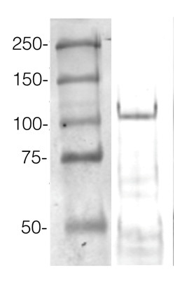
Example of a western blot result obtained with this method, which allows high protein load per well, can be found below.
Agrisera Western Blot protocol and video tutorials
Protocols to work with plant and algal protein extracts
Agrisera Educational Posters Collection
360 µg/well of Arabidopsis thaliana protein extracted by TCA-acetone precipitation from floral tissue and saturated in 8M urea were separated on 15% SDS-PAGE and blotted for 1hour to 0.2 µm nitrocellulose at 100V using wet transfer system. Blots were blocked with 0.5% cold fish gelatin for 1hr at room temp with agitation. Blot was incubated in the primary antibody at a dilution of 1:2500 for an hour at RT with agitation. The blots were washed with 3X 15min TBS-TT at RT with agitation. Blots as incubated in the secondary antibody (DayLight 800) 1:5000 dilution for 30 min. at RT with agitation and washed 1X with TBSTT for 15 min, 1X with TBST for 15min before scanning with the ODyssey IRD scanner.
Courtesy of Dr. Betty Chung and Pawel Baster, University of Cambridge, United Kingdom
BACK to Plant Protocols page - Reviews:
-
This product doesn't have any reviews.
Accessories

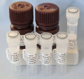
This set contains:
- 4 Anti-AGO antibodies of your choice, chosen from a drop down menu below
- Matching secondary antibody, HRP conjugated, min. 1: 25 000, 1h/RT (AS09 602 - trial)

