1

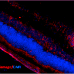
Anti-8-Hydroxyguanosine | DNA/RNA oxidative damage (clone 15A3)
AS10 708-100 | Clonality: monoclonal | Host: Mouse | Reactivity: hydroxy guanosine
- Product Info
-
Sub class: IgG2A Immunogen: 8-hydroxy-guanosine-BSA and – casein conjugates
Host: Mouse Clonality: Monoclonal Purity: Total IgG fraction. Protein G purified. Format: Liquid Quantity: 100 µg Storage: Store lyophilized/reconstituted at -20°C; once reconstituted make aliquots to avoid repeated freeze-thaw cycles. Please remember to spin the tubes briefly prior to opening them to avoid any losses that might occur from material adhering to the cap or sides of the tube. Tested applications: ELISA (ELISA), Immunoaffinity chromatography (IAP), Immunohistochemistry on frozen tissue and paraffin-embedded (IHC-Fr-P) Recommended dilution: The optimal working dilution should be determined by the investigator - Reactivity
-
Confirmed reactivity: Recognizes markers of oxidative damage to DNA (8-hydroxy-2’-deoxyguanosine, 8-hydroxyguanine and 8-hydroxyguanosine) Not reactive in: No confirmed exceptions from predicted reactivity are currently known - Additional Information
-
Additional information: Protein G purified IgG2B in PBS, pH 7,4 with 0,09 % sodium azide and 50 % glycerol at concentration 0,65 mg/ml Additional information (application): Protocol for immunostaining using this antibody can be found here.
- Background
-
Background: Oxidative derivate of guanosine is called 8-Hydroxyguanosine (8OHdG) and is used as a popular biomarker of oxidative stress.
- Product Citations
-
Selected references: Poborilova et al. (2015). DNA hypomethylation concomitant with the overproduction of ROS induced by naphthoquinone juglone on tobacco BY-2 suspension cells. Environmental and Experimental Botany, Volume 113, May 2015, Pages 28–39.
Haigh and Drew (2015). Cavitation during the protein misfolding cyclic amplification (PMCA) method - The trigger for de novo prion generation? Biochem Biophys Res Commun. 2015 Apr 17. pii: S0006-291X(15)00726-3. doi: 10.1016/j.bbrc.2015.04.048.
- Protocols
-
Immunostaining using anti-hydroxy guanosine monoclonal antibody
Tissue Preparation
Anti-hydroxy guanosine antibody monoclonal antibody reacts on both 50 µm frozen tissue sections and paraffin-embedded sections. Tissue should be dissected fresh and fixed in periodate-lysineparaformaldehyde (PLP) at 4°C
overnight.
PLP
Heat 1 L dest. water to 60°C. Add 60 g paraformaldehyde. Add 33 g dibasic NaPO4. Cool to room termperature in a cold water bath. Add 9 g monobasic NaPO4
.
Add 6.45 g Na-m-periodate.
Add 41.1 g lysine (HCl salt).
Filter and dilute to 3 L with dest. water. Adjust pH to 7.6 with 1.0 N NaOH
approx. (20-30 ml).
Tissue prepared for frozen sectioning must be cryoprotected in a 20% glycerol-2% DMSO solution in phosphate buffer for 24-48 hours. Tissue will sink to the bottom of container when fully penetrated. This will eliminate freezing artifact from cutting.Glycerol-DMSO (for 3 L)
2.4 L 0.1 M phosphate buffer
600 ml glycerol
60 ml DMSO0.1 M Phosphate Buffer, pH 7.4 (for 1 L)
1 L dest. water
11 g dibasic NaPO4
3 g monobasic NaPO4
After frozen sectioning, tissue should be stored in phosphate buffer with 0.08% sodium azide.Staining Sections By DAB Procedure
Paraffin-embedded sections must be deparaffinized by sequential immersion in the following for 3 minutes each: xylene (twice), absolute ethanol (twice). Agitate gently in each solution. Proceed with the following procedure:
1. Pretreat sections with a methanolperoxide solution to eliminate endogenous peroxidases.Methanol-Peroxide
100 ml absolute methanol
1 ml 33% H2O2
Incubate sections in methanolperoxide
solution for 30 minutes, room temperature.
2. Wash sections 3 times for 10 minutes each in 0.1 M phosphate buffered saline (PBS)
PBS, pH 7.4 (for 1 L)
1 L dest. water
11 g dibasic NaPO4
3 g monobasic NaPO4
8.5 g NaCl
3. Incubate sections for 1 hour in 10% normal goat serum in PBS.
4. Incubate sections in the primary antibody for 18-24 hours at room temperature. Depending on the nature
of the sample, a shorter incubation time may be used. - Reviews:
-
This product doesn't have any reviews.


