1

Anti-GFP | Green Fluorescence Protein, monoclonal (clone 2G4:F2)
AS21 4696 | Clonality: Monoclonal | Host: Mouse | Reactivity: Green Fluorescence Protein (GFP)
- Product Info
-
Sub class: IgG2a κ-chains Immunogen: Recombinant GFP protein derived from Aequorea victoria, UniProt: P42212
Host: Mouse Clonality: Monoclonal Purity: Purified mouse IgG in PBS pH 7.4 Format: Lyophilized Quantity: 50 µg Reconstitution: For reconstitution add 50 µl, of sterile water Storage: Store lyophilized/reconstituted at -20°C; once reconstituted make aliquots to avoid repeated freeze-thaw cycles, Please, remember to spin tubes briefly prior to opening them to avoid any losses that might occur from lyophilized material adhering to the cap or sides of the tubes Tested applications: Western blot (WB) Recommended dilution: 1: 1000 - 1 : 30 000 (WB) Expected | apparent MW: Depends upon fusion partner - Reactivity
-
Confirmed reactivity: GFP-tagged proteins
- Application Examples
-
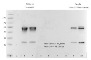
1. Ladder (PageRuler Prestained, 10 à 180 kDa - Thermo)
Samples
2. 20 µg of total protein Col-0 (leaves, control)
3. 20 µg of total Prot-GFP siliques (extract before purification)
4. Purified Prot-GFP siliques - siliques (with Mylteny beads)
5. Purified Elution Prot-GFP siliques (with Mylteny beads) –SDS-buffer
6. Purified Prot-GFP - siliques (with ProteinTech beads)
7. Purified Elution Prot-GFP siliques 6M Urea (with ProteinTech beads)
8. Purified Elution Prot-GFP - SDS-buffer siliques (with ProteinTech beads)
9. 20 µg of total Seeds of A. thaliana Prot-GFP
10. 20 µg of total Seeds of A. thaliana Prot-Venus20 μg/well of total protein extracted freshly from siliques (stade 20dap, 50mg of powder) and imbibed seeds (50 mg starting material). Exact buffer components were Cell Culture Lysis 1X Reagent (Promega - E153A + inhibitor protease cocktail) and denatured with Laemmli buffer (375 mM Tris.HCl, 9% SDS, 50% Glycerol, 0.03% Bromophenol blue) at 95 °C/5 min. Samples were separated on 12 % SDS-PAGE and blotted for 1 h to nitrocellulose (pore size of 0,45 µm), using wet transfer. Blot was blocked with 5 % milk for 2h/RT with agitation. Blot was incubated in the primary antibody at a dilution of 1: 1000 in 5 % milk TBS-T ON/4°C with agitation. The antibody solution was decanted, and the blot was washed 4 times for 5 min in TBS-T at RT with agitation. Blot was incubated with secondary antibody (AS09 627-trial, Rabbit anti-mouse IgG, HRP conjugated, Agrisera) diluted to 1: 10 000 in 5 % milk for 1h/RT with agitation. The blot was washed as above and developed with a following chemiluminescent detection reagent AS16 ECL-S-10, AgriseraSuperBright. Exposure time was 20 seconds.
Courtesy of Dr. Victoria Gomez Roldan,French National Centre for Scientific Research, France
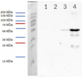
Total proteins were isolated from 4 leaves (5-week-old plants) of Arabidopsis thaliana were ground in 400 μL homogenization buffer or from 15 seedlings (7-day-old) ground in 100 μL homogenization buffer as described by LaMontagne et al (2016). Plant lines were Arabidopsis thaliana Col-0 (ecotype, wild-type) and an overexpressing GFP-tagged ABCG-protein (GFP-ABCG ox). 15 μl of total proteins were denatured at 37°C for 5 min, separated on 8% SDS-PAGE and transferred for 70 min at 55V using a tank transfer system to nitrocellulose membrane (0.45 μm). Blots were blocked with PBS+0.1% Tween 20 (PBS-T)+5 % milk at room temperature (RT) with agitation for 2 hours. Monoclonal primary anti-GFP-antibody (AS21 4696, Agrisera; clone 2G4:F2) was diluted to 1:30,000 and incubated with membrane portion overnight at 4°C with agitation in PBS-T+5 % milk. The primary antibody solution was decanted, and blots were washed 4 times (6 minutes each) in PBS-T at RT with agitation. Blot was incubated with secondary rabbit anti-mouse IgG (H&L) HRP conjugated (AS09 627-trial, Agrisera) diluted to 1: 25,000 for 2 hours, washed as described above (4 times with PBS-T at RT), and developed with chemiluminescent detection reagent ECL Bright (AS16 ECL-N, Agrisera) according to manufacture's recommendations Exposure time was 1 second (1s) on X-ray films. Ponceau Stain of the total proteins and anti-MPK6 detection served as loading control.
Courtesy of Nga Nguyen and Dr. Antje Heese at the University of Missouri-Columbia, Div. of Biochemistry, Interdisciplinary Plant Group (IPG), Columbia MO (USA).
Reference: LaMontagne ED, Collins CA, Peck SC, Heese A. Isolation of Microsomal Membrane Proteins from Arabidopsis thaliana. Curr Protoc Plant Biol. 2016 May;1(1):217-234. doi: 10.1002/cppb.20020. PMID: 31725992. - Background
-
Background: GFP (Green fluorescent protein) was originally identified in photo organs on jellyfish Aequorea victoria. It is a naturally fluorescent protein which emits green light at a maximum wavelength of 509 nm when excited by blue or UV light. It is extensively used in laboratory as a reporter molecule to label and study cellular and subcellular proteins in living cells using a wide range of applications. Antibodies to GFP protein are used in immunoblotting and ELISA. GFP protein has molecular weight of 27 kDa. - Protocols
-
Agrisera Western Blot protocol and video tutorials
Protocols to work with plant and algal protein extracts
Agrisera Educational Poster Collection - Reviews:
-
This product doesn't have any reviews.


